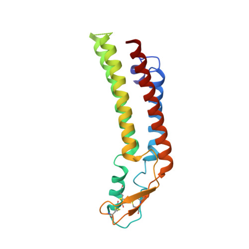
7QEY

human Connexin 26 class 1 hexamer at 90mmHg PCO2, pH7.4


Experimental Data Snapshot

  • Method: ELECTRON MICROSCOPY
  • Resolution: 2.00 Å
  • Aggregation State: PARTICLE 
  • Reconstruction Method: SINGLE PARTICLE 

Starting Model: experimental
View more details

wwPDB Validation   3D Report Full Report


This is version 1.2 of the entry. See complete history


Literature

Conformational changes and CO 2 -induced channel gating in connexin26.

Brotherton, D.H.Savva, C.G.Ragan, T.J.Dale, N.Cameron, A.D.

(2022) Structure 30: 697-706.e4

  • DOI: https://doi.org/10.1016/j.str.2022.02.010
  • Primary Citation of Related Structures:  
    7QEO, 7QEQ, 7QER, 7QES, 7QET, 7QEU, 7QEV, 7QEW, 7QEY

  • PubMed Abstract: 

    Connexins form large-pore channels that function either as dodecameric gap junctions or hexameric hemichannels to allow the regulated movement of small molecules and ions across cell membranes. Opening or closing of the channels is controlled by a variety of stimuli, and dysregulation leads to multiple diseases. An increase in the partial pressure of carbon dioxide (PCO 2 ) has been shown to cause connexin26 (Cx26) gap junctions to close. Here, we use cryoelectron microscopy (cryo-EM) to determine the structure of human Cx26 gap junctions under increasing levels of PCO 2 . We show a correlation between the level of PCO 2 and the size of the aperture of the pore, governed by the N-terminal helices that line the pore. This indicates that CO 2 alone is sufficient to cause conformational changes in the protein. Analysis of the conformational states shows that movements at the N terminus are linked to both subunit rotation and flexing of the transmembrane helices.


  • Organizational Affiliation

    School of Life Sciences, University of Warwick, Gibbet Hill Road, CV4 7AL Coventry, UK.


Macromolecules
Find similar proteins by:  (by identity cutoff)  |  3D Structure
Entity ID: 1
MoleculeChains Sequence LengthOrganismDetailsImage
Gap junction beta-2 protein182Homo sapiensMutation(s): 0 
Gene Names: GJB2
Membrane Entity: Yes 
UniProt & NIH Common Fund Data Resources
Find proteins for P29033 (Homo sapiens)
Explore P29033 
Go to UniProtKB:  P29033
PHAROS:  P29033
GTEx:  ENSG00000165474 
Entity Groups  
Sequence Clusters30% Identity50% Identity70% Identity90% Identity95% Identity100% Identity
UniProt GroupP29033
Sequence Annotations
Expand
  • Reference Sequence
Small Molecules
Ligands 2 Unique
IDChains Name / Formula / InChI Key2D Diagram3D Interactions
PTY
Query on PTY

Download Ideal Coordinates CCD File 
BA [auth K]
H [auth G]
JA [auth L]
M [auth H]
R [auth I]
BA [auth K],
H [auth G],
JA [auth L],
M [auth H],
R [auth I],
W [auth J]
PHOSPHATIDYLETHANOLAMINE
C40 H80 N O8 P
NJGIRBISCGPRPF-KXQOOQHDSA-N
LMT
Query on LMT

Download Ideal Coordinates CCD File 
AA [auth K]
CA [auth K]
DA [auth K]
EA [auth K]
FA [auth L]
AA [auth K],
CA [auth K],
DA [auth K],
EA [auth K],
FA [auth L],
G,
GA [auth L],
HA [auth L],
I [auth G],
IA [auth L],
J [auth G],
K [auth G],
L [auth H],
N [auth H],
O [auth H],
P [auth H],
Q [auth I],
S [auth I],
T [auth I],
U [auth I],
V [auth J],
X [auth J],
Y [auth J],
Z [auth J]
DODECYL-BETA-D-MALTOSIDE
C24 H46 O11
NLEBIOOXCVAHBD-QKMCSOCLSA-N
Experimental Data & Validation

Experimental Data

  • Method: ELECTRON MICROSCOPY
  • Resolution: 2.00 Å
  • Aggregation State: PARTICLE 
  • Reconstruction Method: SINGLE PARTICLE 
EM Software:
TaskSoftware PackageVersion
MODEL REFINEMENTPHENIX1.18.2-3874
RECONSTRUCTIONRELION3.1-beta

Structure Validation

View Full Validation Report


Entry History & Funding Information

Deposition Data


Funding OrganizationLocationGrant Number
Medical Research Council (MRC, United Kingdom)United KingdomMR/P010393/1
Leverhulme TrustUnited KingdomRPG-2015-090

Revision History  (Full details and data files)

  • Version 1.0: 2022-03-30
    Type: Initial release
  • Version 1.1: 2022-11-09
    Changes: Database references
  • Version 1.2: 2024-11-20
    Changes: Data collection, Refinement description, Structure summary