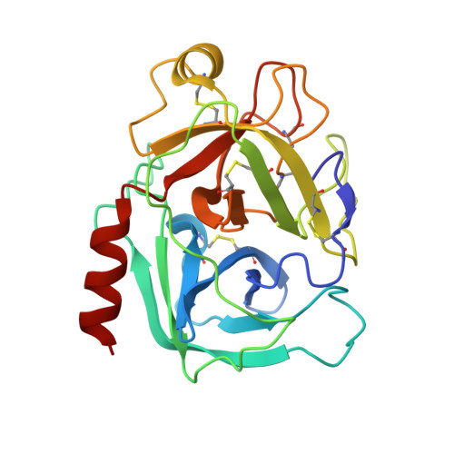
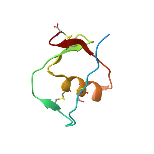
7QE9

Human cationic trypsin (TRY1) complexed with serine protease inhibitor Kazal type 1 N34S (SPINK1 N34S)


Experimental Data Snapshot

  • Method: X-RAY DIFFRACTION
  • Resolution: 2.10 Å
  • R-Value Free: 0.234 
  • R-Value Work: 0.193 

Starting Models: experimental
View more details

wwPDB Validation   3D Report Full Report


This is version 1.3 of the entry. See complete history


Literature

Structural and Biophysical Insights into SPINK1 Bound to Human Cationic Trypsin.

Nagel, F.Palm, G.J.Geist, N.McDonnell, T.C.R.Susemihl, A.Girbardt, B.Mayerle, J.Lerch, M.M.Lammers, M.Delcea, M.

(2022) Int J Mol Sci 23

  • DOI: https://doi.org/10.3390/ijms23073468
  • Primary Citation of Related Structures:  
    7QE8, 7QE9

  • PubMed Abstract: 

    (1) The serine protease inhibitor Kazal type 1 (SPINK1) inhibits trypsin activity in zymogen granules of pancreatic acinar cells. Several mutations in the SPINK1 gene are associated with acute recurrent pancreatitis (ARP) and chronic pancreatitis (CP). The most common variant is SPINK1 p.N34S. Although this mutation was identified two decades ago, the mechanism of action has remained elusive. (2) SPINK1 and human cationic trypsin (TRY1) were expressed in E. coli , and inhibitory activities were determined. Crystals of SPINK1-TRY1 complexes were grown by using the hanging-drop method, and phases were solved by molecular replacement. (3) Both SPINK1 variants show similar inhibitory behavior toward TRY1. The crystal structures are almost identical, with minor differences in the mutated loop. Both complexes show an unexpected rotamer conformation of the His63 residue in TRY1, which is a member of the catalytic triad. (4) The SPINK1 p.N34S mutation does not affect the inhibitory behavior or the overall structure of the protein. Therefore, the pathophysiological mechanism of action of the p.N34S variant cannot be explained mechanistically or structurally at the protein level. The observed histidine conformation is part of a mechanism for SPINK1 that can explain the exceptional proteolytic stability of this inhibitor.


  • Organizational Affiliation

    Biophysical Chemistry, Institute of Biochemistry, University of Greifswald, 17489 Greifswald, Germany.


Macromolecules
Find similar proteins by:  (by identity cutoff)  |  3D Structure
Entity ID: 1
MoleculeChains Sequence LengthOrganismDetailsImage
Trypsin-1
A, B
224Homo sapiensMutation(s): 1 
Gene Names: PRSS1TRP1TRY1TRYP1
EC: 3.4.21.4
UniProt & NIH Common Fund Data Resources
Find proteins for P07477 (Homo sapiens)
Go to UniProtKB:  P07477
PHAROS:  P07477
GTEx:  ENSG00000204983 
Sequence Annotations
Expand
  • Reference Sequence
Find similar proteins by:  (by identity cutoff)  |  3D Structure
Entity ID: 2
MoleculeChains Sequence LengthOrganismDetailsImage
Serine protease inhibitor Kazal-type 1C [auth D],
D [auth C]
61Homo sapiensMutation(s): 1 
Gene Names: SPINK1PSTI
UniProt & NIH Common Fund Data Resources
Find proteins for P00995 (Homo sapiens)
Go to UniProtKB:  P00995
PHAROS:  P00995
GTEx:  ENSG00000164266 
Sequence Annotations
Expand
  • Reference Sequence
Experimental Data & Validation

Experimental Data

  • Method: X-RAY DIFFRACTION
  • Resolution: 2.10 Å
  • R-Value Free: 0.234 
  • R-Value Work: 0.193 
  • Space Group: P 31 2 1
Unit Cell:
Length ( Å )Angle ( ˚ )
a = 76.555α = 90
b = 76.555β = 90
c = 189.719γ = 120
Software Package:
Software NamePurpose
REFMACrefinement
XDSdata reduction
XDSdata scaling
PHASERphasing

Structure Validation

View Full Validation Report


Entry History & Funding Information

Deposition Data


Funding OrganizationLocationGrant Number
European Research Council (ERC)European Union637877
European Union (EU)European UnionESF/14-BM-A55-0047/16

Revision History  (Full details and data files)

  • Version 1.0: 2022-03-30
    Type: Initial release
  • Version 1.1: 2022-04-27
    Changes: Database references
  • Version 1.2: 2024-05-01
    Changes: Data collection, Derived calculations, Refinement description
  • Version 1.3: 2024-10-16
    Changes: Structure summary