


Carboxypeptidase T with (S)-3-phenyllactic acid
Timofeev, V.I., Akparov, V.K., Shevtsov, M.B., Kuranova, I.P.To be published.
Experimental Data Snapshot
Starting Model: experimental
View more details
Entity ID: 1 | |||||
|---|---|---|---|---|---|
| Molecule | Chains | Sequence Length | Organism | Details | Image |
| Carboxypeptidase T | 323 | Thermoactinomyces vulgaris | Mutation(s): 0 Gene Names: cpt EC: 3.4.17.18 | 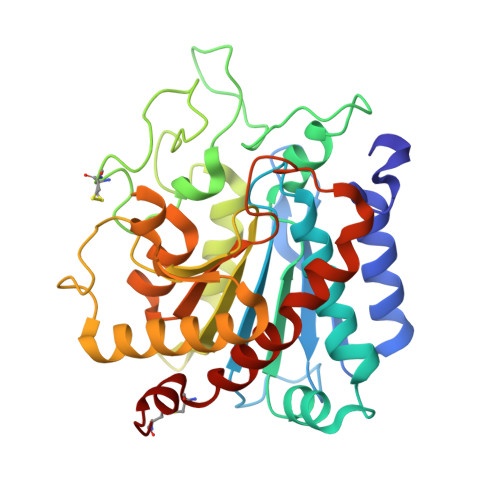 | |
UniProt | |||||
Entity Groups | |||||
| Sequence Clusters | 30% Identity50% Identity70% Identity90% Identity95% Identity100% Identity | ||||
| UniProt Group | P29068 | ||||
Sequence AnnotationsExpand | |||||
Reference Sequence | |||||
| Ligands 4 Unique | |||||
|---|---|---|---|---|---|
| ID | Chains | Name / Formula / InChI Key | 2D Diagram | 3D Interactions | |
| HFA Download:Ideal Coordinates CCD File | C [auth A], D [auth A] | ALPHA-HYDROXY-BETA-PHENYL-PROPIONIC ACID C9 H10 O3 VOXXWSYKYCBWHO-QMMMGPOBSA-N |  | ||
| SO4 Download:Ideal Coordinates CCD File | K [auth A] | SULFATE ION O4 S QAOWNCQODCNURD-UHFFFAOYSA-L |  | ||
| ZN Download:Ideal Coordinates CCD File | B [auth A] | ZINC ION Zn PTFCDOFLOPIGGS-UHFFFAOYSA-N |  | ||
| CA Download:Ideal Coordinates CCD File | E [auth A] F [auth A] G [auth A] H [auth A] I [auth A] | CALCIUM ION Ca BHPQYMZQTOCNFJ-UHFFFAOYSA-N |  | ||
| Length ( Å ) | Angle ( ˚ ) |
|---|---|
| a = 157.328 | α = 90 |
| b = 157.328 | β = 90 |
| c = 104.132 | γ = 120 |
| Software Name | Purpose |
|---|---|
| MOSFLM | data reduction |
| SCALA | data scaling |
| PHASER | phasing |
| REFMAC | refinement |
| PDB_EXTRACT | data extraction |
| Funding Organization | Location | Grant Number |
|---|---|---|
| Not funded | -- |


