7Q53 | pdb_00007q53

Single Particle Cryo-EM structure of photosynthetic A2B2 glyceraldehyde 3-phosphate dehydrogenase from Spinacia oleracia


Experimental Data Snapshot

  • Method: ELECTRON MICROSCOPY
  • Resolution: 6.30 Å
  • Aggregation State: PARTICLE 
  • Reconstruction Method: SINGLE PARTICLE 

Starting Model: experimental
View more details

wwPDB Validation 3D Report Full Report

Validation slider image for 7Q53

This is version 1.2 of the entry. See complete history

Literature

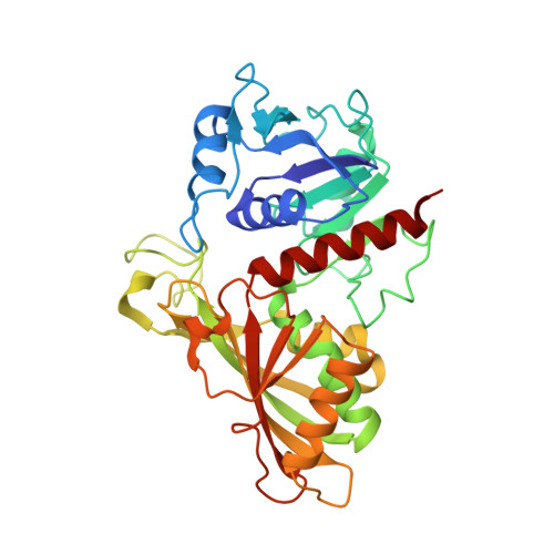
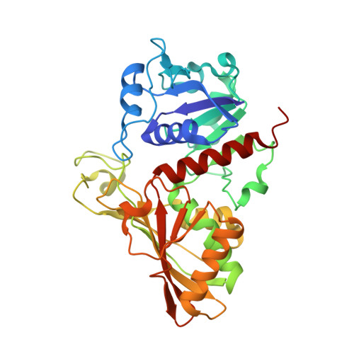
Unravelling the regulation pathway of photosynthetic AB-GAPDH.

Marotta, R.Del Giudice, A.Gurrieri, L.Fanti, S.Swuec, P.Galantini, L.Falini, G.Trost, P.Fermani, S.Sparla, F.

(2022) Acta Crystallogr D Struct Biol 78: 1399-1411

  • DOI: https://doi.org/10.1107/S2059798322010014
  • Primary Citation Related Structures: 
    7Q53, 7Q54, 7Q55, 7Q56, 7Q57

  • PubMed Abstract: 

    Oxygenic phototrophs perform carbon fixation through the Calvin-Benson cycle. Different mechanisms adjust the cycle and the light-harvesting reactions to rapid environmental changes. Photosynthetic glyceraldehyde 3-phosphate dehydrogenase (GAPDH) is a key enzyme in the cycle. In land plants, different photosynthetic GAPDHs exist: the most abundant isoform is formed by A 2 B 2 heterotetramers and the least abundant by A 4 homotetramers. Regardless of the subunit composition, GAPDH is the major consumer of photosynthetic NADPH and its activity is strictly regulated. While A 4 -GAPDH is regulated by CP12, AB-GAPDH is autonomously regulated through the C-terminal extension (CTE) of its B subunits. Reversible inhibition of AB-GAPDH occurs via the oxidation of a cysteine pair located in the CTE and the substitution of NADP(H) with NAD(H) in the cofactor-binding site. These combined conditions lead to a change in the oligomerization state and enzyme inhibition. SEC-SAXS and single-particle cryo-EM analysis were applied to reveal the structural basis of this regulatory mechanism. Both approaches revealed that spinach (A 2 B 2 ) n -GAPDH oligomers with n = 1, 2, 4 and 5 co-exist in a dynamic system. B subunits mediate the contacts between adjacent tetramers in A 4 B 4 and A 8 B 8 oligomers. The CTE of each B subunit penetrates into the active site of a B subunit of the adjacent tetramer, which in turn moves its CTE in the opposite direction, effectively preventing the binding of the substrate 1,3-bisphosphoglycerate in the B subunits. The whole mechanism is made possible, and eventually controlled, by pyridine nucleotides. In fact, NAD(H), by removing NADP(H) from A subunits, allows the entrance of the CTE into the active site of the B subunit, hence stabilizing inhibited oligomers.


  • Organizational Affiliation
    • Electron Microscopy Facility (EMF), Italian Institute of Technology (IIT), 16163 Genova, Italy.

Macromolecule Content 

  • Total Structure Weight: 147.74 kDa 
  • Atom Count: 10,362 
  • Modeled Residue Count: 1,352 
  • Deposited Residue Count: 1,352 
  • Unique protein chains: 2

Macromolecules

Find similar proteins by:|  3D Structure
Entity ID: 1
MoleculeChains  Sequence LengthOrganismDetailsImage
Glyceraldehyde-3-phosphate dehydrogenase B, chloroplasticA [auth O],
C [auth Q]
339Spinacia oleraceaMutation(s): 0 
EC: 1.2.1.13
UniProt
Find proteins for P12860 (Spinacia oleracea)
Explore P12860 
Go to UniProtKB:  P12860
Entity Groups
Sequence Clusters30% Identity50% Identity70% Identity90% Identity95% Identity100% Identity
UniProt GroupP12860
Sequence Annotations
Expand
Reference Sequence
Find similar proteins by:|  3D Structure
Entity ID: 2
MoleculeChains  Sequence LengthOrganismDetailsImage
Glyceraldehyde-3-phosphate dehydrogenase A, chloroplastic,Glyceraldehyde-3-phosphate dehydrogenase A, chloroplastic,Glyceraldehyde-3-phosphate dehydrogenase A, chloroplastic,Glyceraldehyde-3-phosphate dehydrogenase A, chloroplasticB [auth P],
D [auth R]
337Spinacia oleraceaMutation(s): 0 
EC: 1.2.1.13
UniProt
Find proteins for P19866 (Spinacia oleracea)
Explore P19866 
Go to UniProtKB:  P19866
Entity Groups
Sequence Clusters30% Identity50% Identity70% Identity90% Identity95% Identity100% Identity
UniProt GroupP19866
Sequence Annotations
Expand
Reference Sequence

Experimental Data & Validation

Experimental Data

  • Method: ELECTRON MICROSCOPY
  • Resolution: 6.30 Å
  • Aggregation State: PARTICLE 
  • Reconstruction Method: SINGLE PARTICLE 
EM Software:
TaskSoftware PackageVersion
MODEL REFINEMENTPHENIX

Structure Validation

View Full Validation Report


Entry History 

& Funding Information

Deposition Data


Funding OrganizationLocationGrant Number
Grenoble Instruct-ERIC Center (ISBG)FrancePID 1829

Revision History  (Full details and data files)

  • Version 1.0: 2022-11-16
    Type: Initial release
  • Version 1.1: 2024-07-17
    Changes: Data collection, Refinement description
  • Version 1.2: 2025-07-02
    Changes: Data collection, Structure summary