7Q4T

Structure of the Pseudomonas aeruginosa bacteriophage JG004 endolysin Pae87 bound to a peptidoglycan fragment.


Experimental Data Snapshot

  • Method: X-RAY DIFFRACTION
  • Resolution: 1.27 Å
  • R-Value Free: 0.161 
  • R-Value Work: 0.130 

Starting Model: experimental
View more details

wwPDB Validation   3D Report Full Report


This is version 1.2 of the entry. See complete history


Literature

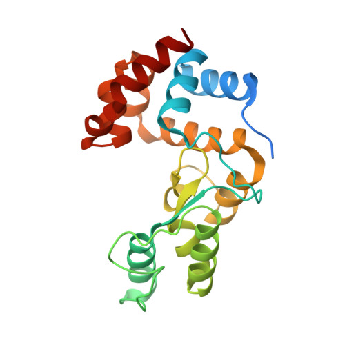
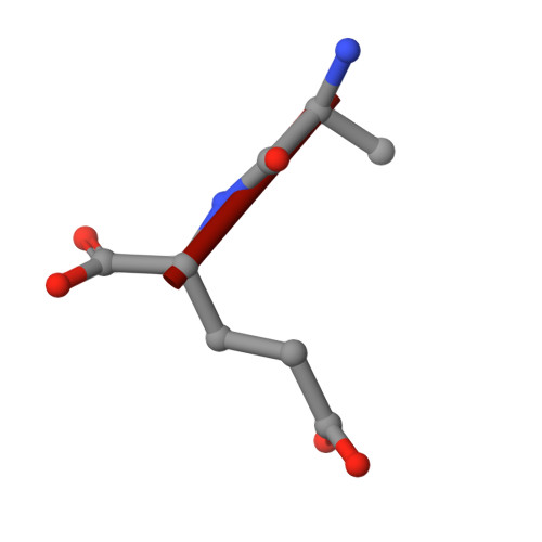
Monomodular Pseudomonas aeruginosa phage JG004 lysozyme (Pae87) contains a bacterial surface-active antimicrobial peptide-like region and a possible substrate-binding subdomain.

Vazquez, R.Seoane-Blanco, M.Rivero-Buceta, V.Ruiz, S.van Raaij, M.J.Garcia, P.

(2022) Acta Crystallogr D Struct Biol 78: 435-454

  • DOI: https://doi.org/10.1107/S2059798322000936
  • Primary Citation of Related Structures:  
    7Q4S, 7Q4T

  • PubMed Abstract: 

    Phage lysins are a source of novel antimicrobials to tackle the bacterial antibiotic-resistance crisis. The engineering of phage lysins is being explored as a game-changing technological strategy to introduce a more precise approach in the way in which antimicrobial therapy is applied. Such engineering efforts will benefit from a better understanding of lysin structure and function. In this work, the antimicrobial activity of the endolysin from Pseudomonas aeruginosa phage JG004, termed Pae87, has been characterized. This lysin had previously been identified as an antimicrobial agent candidate that is able to interact with the Gram-negative surface and disrupt it. Further evidence is provided here based on a structural and biochemical study. A high-resolution crystal structure of Pae87 complexed with a peptidoglycan fragment showed a separate substrate-binding region within the catalytic domain, 18 Å away from the catalytic site and located on the opposite side of the lysin molecule. This substrate-binding region was conserved among phylogenetically related lysins lacking an additional cell-wall-binding domain, but not among those containing such a module. Two glutamic acids were identified to be relevant for the peptidoglycan-degradation activity, although the antimicrobial activity of Pae87 was seemingly unrelated. In contrast, an antimicrobial peptide-like region within the Pae87 C-terminus, named P87, was found to be able to actively disturb the outer membrane and display antibacterial activity by itself. Therefore, an antimicrobial mechanism for Pae87 is proposed in which the P87 peptide plays the role of binding to the outer membrane and disrupting the cell-wall function, either with or without the participation of the catalytic activity of Pae87.


  • Organizational Affiliation

    Microbial and Plant Biotechnology Department, Centro de Investigaciones Biológicas Margarita Salas (CIB-CSIC), Madrid, Spain.


Macromolecules
Find similar proteins by:  (by identity cutoff)  |  3D Structure
Entity ID: 1
MoleculeChains Sequence LengthOrganismDetailsImage
EndolysinA [auth AAA]206Pseudomonas phage JG004Mutation(s): 0 
Gene Names: PJG4_087
UniProt
Find proteins for F4YDQ3 (Pseudomonas phage JG004)
Explore F4YDQ3 
Go to UniProtKB:  F4YDQ3
Entity Groups  
Sequence Clusters30% Identity50% Identity70% Identity90% Identity95% Identity100% Identity
UniProt GroupF4YDQ3
Sequence Annotations
Expand
  • Reference Sequence

Find similar proteins by:  Sequence   |   3D Structure  

Entity ID: 2
MoleculeChains Sequence LengthOrganismDetailsImage
ALA-DGLB [auth LbL]2Pseudomonas aeruginosa PAO1Mutation(s): 0 
Glycosylation
Glycosylation Sites: 1
Sequence Annotations
Expand
  • Reference Sequence
Oligosaccharides

Help

Entity ID: 3
MoleculeChains Length2D Diagram Glycosylation3D Interactions
2-acetamido-2-deoxy-beta-D-glucopyranose-(1-4)-N-acetyl-alpha-muramic acidC [auth LLL]2N/A
Glycosylation Resources
GlyTouCan:  G88499BX
GlyCosmos:  G88499BX
Small Molecules
Ligands 2 Unique
IDChains Name / Formula / InChI Key2D Diagram3D Interactions
PEG
Query on PEG

Download Ideal Coordinates CCD File 
D [auth AAA],
E [auth AAA]
DI(HYDROXYETHYL)ETHER
C4 H10 O3
MTHSVFCYNBDYFN-UHFFFAOYSA-N
EDO
Query on EDO

Download Ideal Coordinates CCD File 
F [auth AAA]
G [auth AAA]
H [auth AAA]
I [auth AAA]
J [auth AAA]
F [auth AAA],
G [auth AAA],
H [auth AAA],
I [auth AAA],
J [auth AAA],
K [auth AAA],
L [auth AAA],
M [auth AAA],
N [auth AAA],
O [auth AAA],
P [auth AAA],
Q [auth AAA],
R [auth LbL]
1,2-ETHANEDIOL
C2 H6 O2
LYCAIKOWRPUZTN-UHFFFAOYSA-N
Experimental Data & Validation

Experimental Data

  • Method: X-RAY DIFFRACTION
  • Resolution: 1.27 Å
  • R-Value Free: 0.161 
  • R-Value Work: 0.130 
  • Space Group: P 21 21 21
Unit Cell:
Length ( Å )Angle ( ˚ )
a = 43.581α = 90
b = 61.173β = 90
c = 69.21γ = 90
Software Package:
Software NamePurpose
REFMACrefinement
XDSdata reduction
Aimlessdata scaling
MOLREPphasing

Structure Validation

View Full Validation Report


Entry History & Funding Information

Deposition Data


Funding OrganizationLocationGrant Number
Ministerio de Ciencia e Innovacion (MCIN)SpainBFU2017-82207-P

Revision History  (Full details and data files)

  • Version 1.0: 2022-02-23
    Type: Initial release
  • Version 1.1: 2022-04-13
    Changes: Database references
  • Version 1.2: 2024-01-31
    Changes: Data collection, Derived calculations, Refinement description