7Q4M

Type II beta-amyloid 42 Filaments from Human Brain


Experimental Data Snapshot

  • Method: ELECTRON MICROSCOPY
  • Resolution: 2.80 Å
  • Aggregation State: FILAMENT 
  • Reconstruction Method: HELICAL 

wwPDB Validation   3D Report Full Report


This is version 1.2 of the entry. See complete history


Literature

Cryo-EM structures of amyloid-beta 42 filaments from human brains.

Yang, Y.Arseni, D.Zhang, W.Huang, M.Lovestam, S.Schweighauser, M.Kotecha, A.Murzin, A.G.Peak-Chew, S.Y.Macdonald, J.Lavenir, I.Garringer, H.J.Gelpi, E.Newell, K.L.Kovacs, G.G.Vidal, R.Ghetti, B.Ryskeldi-Falcon, B.Scheres, S.H.W.Goedert, M.

(2022) Science 375: 167-172

  • DOI: https://doi.org/10.1126/science.abm7285
  • Primary Citation of Related Structures:  
    7Q4B, 7Q4M

  • PubMed Abstract: 

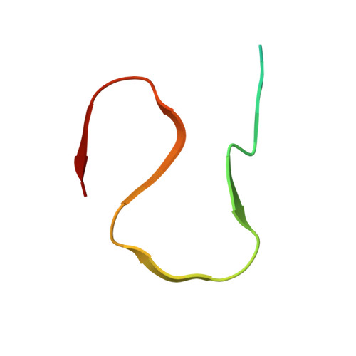
    Filament assembly of amyloid-β peptides ending at residue 42 (Aβ42) is a central event in Alzheimer’s disease. Here, we report the cryo–electron microscopy (cryo-EM) structures of Aβ42 filaments from human brains. Two structurally related S-shaped protofilament folds give rise to two types of filaments. Type I filaments were found mostly in the brains of individuals with sporadic Alzheimer’s disease, and type II filaments were found in individuals with familial Alzheimer’s disease and other conditions. The structures of Aβ42 filaments from the brain differ from those of filaments assembled in vitro. By contrast, in App NL-F knock-in mice, Aβ42 deposits were made of type II filaments. Knowledge of Aβ42 filament structures from human brains may lead to the development of inhibitors of assembly and improved imaging agents.


  • Organizational Affiliation

    Medical Research Council Laboratory of Molecular Biology, Cambridge, UK.


Macromolecules
Find similar proteins by:  (by identity cutoff)  |  3D Structure
Entity ID: 1
MoleculeChains Sequence LengthOrganismDetailsImage
Amyloid-beta precursor protein
A, B, C, D, E
A, B, C, D, E, F, G, H, I, J
42Homo sapiensMutation(s): 0 
UniProt & NIH Common Fund Data Resources
Find proteins for P05067 (Homo sapiens)
Explore P05067 
Go to UniProtKB:  P05067
PHAROS:  P05067
GTEx:  ENSG00000142192 
Entity Groups  
Sequence Clusters30% Identity50% Identity70% Identity90% Identity95% Identity100% Identity
UniProt GroupP05067
Sequence Annotations
Expand
  • Reference Sequence
Experimental Data & Validation

Experimental Data

  • Method: ELECTRON MICROSCOPY
  • Resolution: 2.80 Å
  • Aggregation State: FILAMENT 
  • Reconstruction Method: HELICAL 

Structure Validation

View Full Validation Report


Entry History & Funding Information

Deposition Data


Funding OrganizationLocationGrant Number
Medical Research Council (MRC, United Kingdom)United StatesMC_UP_1201/25
Medical Research Council (MRC, United Kingdom)United StatesMC_UP_A025_1013
Medical Research Council (MRC, United Kingdom)United StatesMC_U105184291
National Institutes of Health/National Institute on Aging (NIH/NIA)United StatesP30-AG010133
National Institutes of Health/National Institute of Neurological Disorders and Stroke (NIH/NINDS)United StatesU01-NS110437
National Institutes of Health/National Institute on Aging (NIH/NIA)United StatesRF1-AG071177

Revision History  (Full details and data files)

  • Version 1.0: 2021-11-24
    Type: Initial release
  • Version 1.1: 2022-01-26
    Changes: Database references
  • Version 1.2: 2023-03-08
    Changes: Structure summary