7Q3J | pdb_00007q3j

Computationally designed thioredoxin subjected to stability optimizing mutations.


Experimental Data Snapshot

  • Method: X-RAY DIFFRACTION
  • Resolution: 1.90 Å
  • R-Value Free: 
    0.239 (Depositor), 0.241 (DCC) 
  • R-Value Work: 
    0.192 (Depositor), 0.193 (DCC) 
  • R-Value Observed: 
    0.195 (Depositor) 

Starting Model: experimental
View more details

wwPDB Validation 3D Report Full Report

Validation slider image for 7Q3J

This is version 1.2 of the entry. See complete history

Literature

Increasing protein stability by inferring substitution effects from high-throughput experiments.

Norrild, R.K.Johansson, K.E.O'Shea, C.Morth, J.P.Lindorff-Larsen, K.Winther, J.R.

(2022) Cell Rep Methods 2: 100333-100333

  • DOI: https://doi.org/10.1016/j.crmeth.2022.100333
  • Primary Citation Related Structures: 
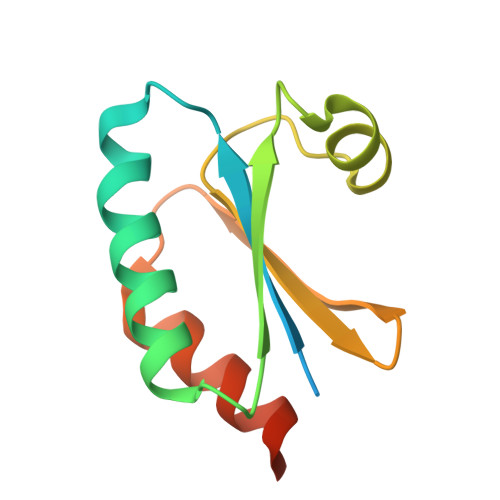
    7Q3J, 7Q3K

  • PubMed Abstract: 

    We apply a computational model, global multi-mutant analysis (GMMA), to inform on effects of most amino acid substitutions from a randomly mutated gene library. Using a high mutation frequency, the method can determine mutations that increase the stability of even very stable proteins for which conventional selection systems have reached their limit. As a demonstration of this, we screened a mutant library of a highly stable and computationally redesigned model protein using an in vivo genetic sensor for folding and assigned a stability effect to 374 of 912 possible single amino acid substitutions. Combining the top 9 substitutions increased the unfolding energy 47 to 69 kJ/mol in a single engineering step. Crystal structures of stabilized variants showed small perturbations in helices 1 and 2, which rendered them closer in structure to the redesign template. This case study illustrates the capability of the method, which is applicable to any screen for protein function.


  • Organizational Affiliation
    • Linderstrøm-Lang Centre for Protein Science, Department of Biology, University of Copenhagen, 2200 Copenhagen N, Denmark.

Macromolecule Content 

  • Total Structure Weight: 26.94 kDa 
  • Atom Count: 1,532 
  • Modeled Residue Count: 176 
  • Deposited Residue Count: 228 
  • Unique protein chains: 1

Macromolecules

Find similar proteins by:|  3D Structure
Entity ID: 1
MoleculeChains  Sequence LengthOrganismDetailsImage
MM9
A, B
114synthetic constructMutation(s): 0 

Experimental Data & Validation

Experimental Data

  • Method: X-RAY DIFFRACTION
  • Resolution: 1.90 Å
  • R-Value Free:  0.239 (Depositor), 0.241 (DCC) 
  • R-Value Work:  0.192 (Depositor), 0.193 (DCC) 
  • R-Value Observed: 0.195 (Depositor) 
Space Group: C 1 2 1
Unit Cell:
Length ( Å )Angle ( ˚ )
a = 58.89α = 90
b = 45.656β = 92.155
c = 72.882γ = 90
Software Package:
Software NamePurpose
PHENIXrefinement
PHENIXrefinement
xia2data reduction
xia2data scaling
PHASERphasing

Structure Validation

View Full Validation Report


Entry History 

& Funding Information

Deposition Data


Funding OrganizationLocationGrant Number
Danish Council for Independent ResearchDenmark--

Revision History  (Full details and data files)

  • Version 1.0: 2022-11-16
    Type: Initial release
  • Version 1.1: 2023-03-01
    Changes: Database references
  • Version 1.2: 2024-02-07
    Changes: Data collection, Refinement description