7Q1F

CPAP:TUBULIN:IE5 ALPHAREP COMPLEX


Experimental Data Snapshot

  • Method: X-RAY DIFFRACTION
  • Resolution: 2.35 Å
  • R-Value Free: 0.220 
  • R-Value Work: 0.188 
  • R-Value Observed: 0.190 

wwPDB Validation   3D Report Full Report


Ligand Structure Quality Assessment 


This is version 1.2 of the entry. See complete history


Literature

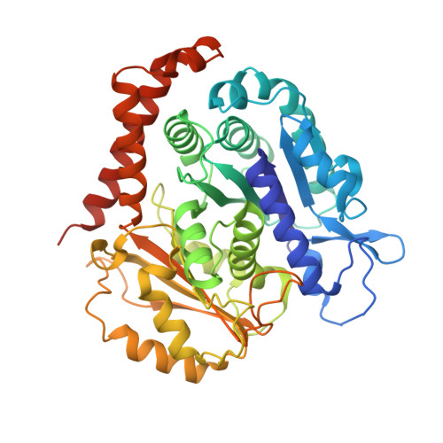
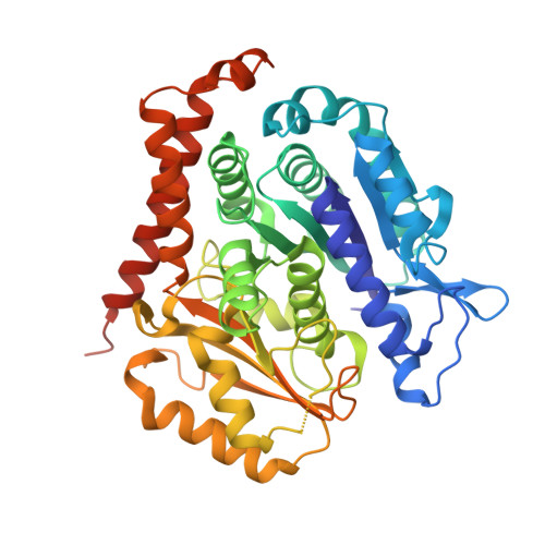
Structural convergence for tubulin binding of CPAP and vinca domain microtubule inhibitors.

Campanacci, V.Urvoas, A.Ammar Khodja, L.Aumont-Nicaise, M.Noiray, M.Lachkar, S.Curmi, P.A.Minard, P.Gigant, B.

(2022) Proc Natl Acad Sci U S A 119: e2120098119-e2120098119

  • DOI: https://doi.org/10.1073/pnas.2120098119
  • Primary Citation of Related Structures:  
    7Q1E, 7Q1F, 7Z0F, 7Z0G

  • PubMed Abstract: 

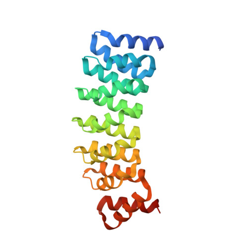
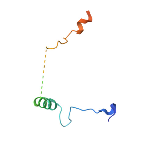
    Microtubule dynamics is regulated by various cellular proteins and perturbed by small-molecule compounds. To what extent the mechanism of the former resembles that of the latter is an open question. We report here structures of tubulin bound to the PN2-3 domain of CPAP, a protein controlling the length of the centrioles. We show that an α-helix of the PN2-3 N-terminal region binds and caps the longitudinal surface of the tubulin β subunit. Moreover, a PN2-3 N-terminal stretch lies in a β-tubulin site also targeted by fungal and bacterial peptide-like inhibitors of the vinca domain, sharing a very similar binding mode with these compounds. Therefore, our results identify several characteristic features of cellular partners that bind to this site and highlight a structural convergence of CPAP with small-molecule inhibitors of microtubule assembly.


  • Organizational Affiliation

    Université Paris-Saclay, CEA, CNRS, Institute for Integrative Biology of the Cell (I2BC), 91198, Gif-sur-Yvette, France.


Macromolecules
Find similar proteins by:  (by identity cutoff)  |  3D Structure
Entity ID: 1
MoleculeChains Sequence LengthOrganismDetailsImage
Tubulin alpha chainA,
F [auth R]
451Ovis ariesMutation(s): 0 
Entity Groups  
Sequence Clusters30% Identity50% Identity70% Identity90% Identity95% Identity100% Identity
Sequence Annotations
Expand
  • Reference Sequence
Find similar proteins by:  (by identity cutoff)  |  3D Structure
Entity ID: 2
MoleculeChains Sequence LengthOrganismDetailsImage
Tubulin beta chainB,
G [auth S]
445Ovis ariesMutation(s): 0 
Entity Groups  
Sequence Clusters30% Identity50% Identity70% Identity90% Identity95% Identity100% Identity
Sequence Annotations
Expand
  • Reference Sequence
Find similar proteins by:  (by identity cutoff)  |  3D Structure
Entity ID: 3
MoleculeChains Sequence LengthOrganismDetailsImage
IE5 ALPHAREPC,
D,
H [auth T],
I [auth U]
232synthetic constructMutation(s): 0 
Entity Groups  
Sequence Clusters30% Identity50% Identity70% Identity90% Identity95% Identity100% Identity
Sequence Annotations
Expand
  • Reference Sequence
Find similar proteins by:  (by identity cutoff)  |  3D Structure
Entity ID: 4
MoleculeChains Sequence LengthOrganismDetailsImage
Centromere protein JE [auth P],
J [auth V]
75Homo sapiensMutation(s): 0 
Gene Names: CENPJCPAPLAPLIP1
UniProt & NIH Common Fund Data Resources
Find proteins for Q9HC77 (Homo sapiens)
Explore Q9HC77 
Go to UniProtKB:  Q9HC77
PHAROS:  Q9HC77
GTEx:  ENSG00000151849 
Entity Groups  
Sequence Clusters30% Identity50% Identity70% Identity90% Identity95% Identity100% Identity
UniProt GroupQ9HC77
Sequence Annotations
Expand
  • Reference Sequence
Small Molecules
Ligands 6 Unique
IDChains Name / Formula / InChI Key2D Diagram3D Interactions
GTP
Query on GTP

Download Ideal Coordinates CCD File 
K [auth A],
R
GUANOSINE-5'-TRIPHOSPHATE
C10 H16 N5 O14 P3
XKMLYUALXHKNFT-UUOKFMHZSA-N
GDP
Query on GDP

Download Ideal Coordinates CCD File 
N [auth B],
U [auth S]
GUANOSINE-5'-DIPHOSPHATE
C10 H15 N5 O11 P2
QGWNDRXFNXRZMB-UUOKFMHZSA-N
MES
Query on MES

Download Ideal Coordinates CCD File 
O [auth B],
P [auth B],
V [auth S],
W [auth S]
2-(N-MORPHOLINO)-ETHANESULFONIC ACID
C6 H13 N O4 S
SXGZJKUKBWWHRA-UHFFFAOYSA-N
FLC
Query on FLC

Download Ideal Coordinates CCD File 
Q [auth D]CITRATE ANION
C6 H5 O7
KRKNYBCHXYNGOX-UHFFFAOYSA-K
SO4
Query on SO4

Download Ideal Coordinates CCD File 
M [auth A],
T [auth R]
SULFATE ION
O4 S
QAOWNCQODCNURD-UHFFFAOYSA-L
MG
Query on MG

Download Ideal Coordinates CCD File 
L [auth A],
S [auth R]
MAGNESIUM ION
Mg
JLVVSXFLKOJNIY-UHFFFAOYSA-N
Experimental Data & Validation

Experimental Data

  • Method: X-RAY DIFFRACTION
  • Resolution: 2.35 Å
  • R-Value Free: 0.220 
  • R-Value Work: 0.188 
  • R-Value Observed: 0.190 
  • Space Group: P 1 21 1
Unit Cell:
Length ( Å )Angle ( ˚ )
a = 89.088α = 90
b = 215.864β = 109.61
c = 95.536γ = 90
Software Package:
Software NamePurpose
BUSTERrefinement
XDSdata reduction
STARANISOdata scaling
PHASERphasing

Structure Validation

View Full Validation Report


Ligand Structure Quality Assessment 


Entry History & Funding Information

Deposition Data


Funding OrganizationLocationGrant Number
Fondation ARCFrance--
Centre National de la Recherche Scientifique (CNRS)France--

Revision History  (Full details and data files)

  • Version 1.0: 2022-04-20
    Type: Initial release
  • Version 1.1: 2022-05-18
    Changes: Database references
  • Version 1.2: 2024-01-31
    Changes: Data collection, Refinement description