7PSQ | pdb_00007psq

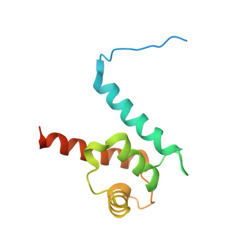
Crystal structure of S100A4 labeled with NU074381b.


Experimental Data Snapshot

  • Method: X-RAY DIFFRACTION
  • Resolution: 1.91 Å
  • R-Value Free: 
    0.236 (Depositor), 0.243 (DCC) 
  • R-Value Work: 
    0.205 (Depositor), 0.213 (DCC) 
  • R-Value Observed: 
    0.206 (Depositor) 

Starting Model: experimental
View more details

wwPDB Validation 3D Report Full Report

Validation slider image for 7PSQ

Ligand Structure Quality Assessment 


This is version 1.2 of the entry. See complete history

Literature

Macromolecule Content 

  • Total Structure Weight: 58.52 kDa 
  • Atom Count: 3,426 
  • Modeled Residue Count: 410 
  • Deposited Residue Count: 492 
  • Unique protein chains: 1

Macromolecules

Find similar proteins by:|  3D Structure
Entity ID: 1
MoleculeChains  Sequence LengthOrganismDetailsImage
Protein S100-A4A,
B [auth C],
C [auth E],
D [auth G]
123Homo sapiensMutation(s): 0 
Gene Names: S100A4CAPLMTS1
UniProt & NIH Common Fund Data Resources
Find proteins for P26447 (Homo sapiens)
Explore P26447 
Go to UniProtKB:  P26447
PHAROS:  P26447
GTEx:  ENSG00000196154 
Entity Groups
Sequence Clusters30% Identity50% Identity70% Identity90% Identity95% Identity100% Identity
UniProt GroupP26447
Sequence Annotations
Expand
Reference Sequence

Small Molecules

Ligands 3 Unique
IDChains Name / Formula / InChI Key2D Diagram3D Interactions
82Q
(Subject of Investigation/LOI)

Query on 82Q


Download:Ideal Coordinates CCD File
L [auth E],
O [auth G]
(2~{R},4~{R})-1-ethanoyl-~{N}-naphthalen-1-yl-4-phenyl-pyrrolidine-2-carboxamide
C23 H22 N2 O2
HXZFWDRYUUGFOU-SIKLNZKXSA-N
EDO

Query on EDO


Download:Ideal Coordinates CCD File
G [auth A],
J [auth C],
K [auth C]
1,2-ETHANEDIOL
C2 H6 O2
LYCAIKOWRPUZTN-UHFFFAOYSA-N
CA

Query on CA


Download:Ideal Coordinates CCD File
E [auth A]
F [auth A]
H [auth C]
I [auth C]
M [auth E]
E [auth A],
F [auth A],
H [auth C],
I [auth C],
M [auth E],
N [auth E],
P [auth G],
Q [auth G],
R [auth G]
CALCIUM ION
Ca
BHPQYMZQTOCNFJ-UHFFFAOYSA-N

Experimental Data & Validation

Experimental Data

  • Method: X-RAY DIFFRACTION
  • Resolution: 1.91 Å
  • R-Value Free:  0.236 (Depositor), 0.243 (DCC) 
  • R-Value Work:  0.205 (Depositor), 0.213 (DCC) 
  • R-Value Observed: 0.206 (Depositor) 
Space Group: P 2 21 21
Unit Cell:
Length ( Å )Angle ( ˚ )
a = 64.913α = 90
b = 70.404β = 90
c = 107.64γ = 90
Software Package:
Software NamePurpose
PHENIXrefinement
PHASERphasing

Structure Validation

View Full Validation Report


Ligand Structure Quality Assessment 


Entry History 

& Funding Information

Deposition Data


Funding OrganizationLocationGrant Number
Innovative Medicines InitiativeSwitzerland115766

Revision History  (Full details and data files)

  • Version 1.0: 2022-10-05
    Type: Initial release
  • Version 1.1: 2024-01-31
    Changes: Data collection, Refinement description
  • Version 1.2: 2024-11-13
    Changes: Structure summary