7PRV | pdb_00007prv

The glucocorticoid receptor in complex with fluticasone furoate, a PGC1a coactivator fragment and sgk 23bp


Experimental Data Snapshot

  • Method: X-RAY DIFFRACTION
  • Resolution: 2.70 Å
  • R-Value Free: 
    0.253 (Depositor), 0.254 (DCC) 
  • R-Value Work: 
    0.214 (Depositor), 0.216 (DCC) 
  • R-Value Observed: 
    0.216 (Depositor) 

Starting Models: experimental
View more details

wwPDB Validation 3D Report Full Report

Validation slider image for 7PRV

Ligand Structure Quality Assessment 


This is version 1.3 of the entry. See complete history

Literature

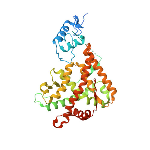
Quaternary glucocorticoid receptor structure highlights allosteric interdomain communication.

Postel, S.Wissler, L.Johansson, C.A.Gunnarsson, A.Gordon, E.Collins, B.Castaldo, M.Kohler, C.Oling, D.Johansson, P.Froderberg Roth, L.Beinsteiner, B.Dainty, I.Delaney, S.Klaholz, B.P.Billas, I.M.L.Edman, K.

(2023) Nat Struct Mol Biol 30: 286-295

  • DOI: https://doi.org/10.1038/s41594-022-00914-4
  • Primary Citation Related Structures: 
    7PRV, 7PRW, 7PRX

  • PubMed Abstract: 

    The glucocorticoid receptor (GR) is a ligand-activated transcription factor that binds DNA and assembles co-regulator complexes to regulate gene transcription. GR agonists are widely prescribed to people with inflammatory and autoimmune diseases. Here we present high-resolution, multidomain structures of GR in complex with ligand, DNA and co-regulator peptide. The structures reveal how the receptor forms an asymmetric dimer on the DNA and provide a detailed view of the domain interactions within and across the two monomers. Hydrogen-deuterium exchange and DNA-binding experiments demonstrate that ligand-dependent structural changes are communicated across the different domains in the full-length receptor. This study demonstrates how GR forms a distinct architecture on DNA and how signal transmission can be modulated by the ligand pharmacophore, provides a platform to build a new level of understanding of how receptor modifications can drive disease progression and offers key insight for future drug design.


  • Organizational Affiliation
    • Mechanistic & Structural Biology, Discovery Sciences, R&D, AstraZeneca, Gothenburg, Sweden.

Macromolecule Content 

  • Total Structure Weight: 106.89 kDa 
  • Atom Count: 6,278 
  • Modeled Residue Count: 698 
  • Deposited Residue Count: 853 
  • Unique protein chains: 2
  • Unique nucleic acid chains: 2

Macromolecules


Find similar proteins by:|  3D Structure
Entity ID: 1
MoleculeChains  Sequence LengthOrganismDetailsImage
Glucocorticoid receptor
A, B
393Homo sapiensMutation(s): 5 
Gene Names: NR3C1GRL
UniProt & NIH Common Fund Data Resources
Find proteins for P04150 (Homo sapiens)
Explore P04150 
Go to UniProtKB:  P04150
PHAROS:  P04150
GTEx:  ENSG00000113580 
Entity Groups
Sequence Clusters30% Identity50% Identity70% Identity90% Identity95% Identity100% Identity
UniProt GroupP04150
Sequence Annotations
Expand
Reference Sequence
Find similar proteins by:  Sequence   |   3D Structure  
Entity ID: 4
MoleculeChains  Sequence LengthOrganismDetailsImage
Peroxisome proliferator-activated receptor gamma coactivator 1-alphaE [auth F]21Homo sapiensMutation(s): 0 
UniProt & NIH Common Fund Data Resources
Find proteins for Q9UBK2 (Homo sapiens)
Explore Q9UBK2 
Go to UniProtKB:  Q9UBK2
PHAROS:  Q9UBK2
GTEx:  ENSG00000109819 
Entity Groups
Sequence Clusters30% Identity50% Identity70% Identity90% Identity95% Identity100% Identity
UniProt GroupQ9UBK2
Sequence Annotations
Expand
Reference Sequence
Find similar nucleic acids by:  Sequence
Entity ID: 2
MoleculeChains LengthOrganismImage
DNA (5'-D(*TP*CP*GP*AP*CP*GP*GP*AP*CP*AP*AP*AP*AP*TP*GP*TP*TP*CP*TP*GP*TP*A)-3')23Homo sapiens
Sequence Annotations
Expand
Reference Sequence
Find similar nucleic acids by:  Sequence
Entity ID: 3
MoleculeChains LengthOrganismImage
DNA (5'-D(*TP*AP*CP*AP*GP*AP*AP*CP*AP*TP*TP*TP*TP*GP*TP*CP*CP*GP*TP*CP*GP*A)-3')23Homo sapiens
Sequence Annotations
Expand
Reference Sequence

Small Molecules

Ligands 3 Unique
IDChains Name / Formula / InChI Key2D Diagram3D Interactions
GW6
(Subject of Investigation/LOI)

Query on GW6


Download:Ideal Coordinates CCD File
J [auth A],
N [auth B]
(6alpha,11alpha,14beta,16alpha,17alpha)-6,9-difluoro-17-{[(fluoromethyl)sulfanyl]carbonyl}-11-hydroxy-16-methyl-3-oxoan drosta-1,4-dien-17-yl furan-2-carboxylate
C27 H29 F3 O6 S
XTULMSXFIHGYFS-VLSRWLAYSA-N
ZN

Query on ZN


Download:Ideal Coordinates CCD File
F [auth A],
G [auth A],
L [auth B],
M [auth B]
ZINC ION
Zn
PTFCDOFLOPIGGS-UHFFFAOYSA-N
EDO

Query on EDO


Download:Ideal Coordinates CCD File
H [auth A],
I [auth A],
K [auth A]
1,2-ETHANEDIOL
C2 H6 O2
LYCAIKOWRPUZTN-UHFFFAOYSA-N

Experimental Data & Validation

Experimental Data

  • Method: X-RAY DIFFRACTION
  • Resolution: 2.70 Å
  • R-Value Free:  0.253 (Depositor), 0.254 (DCC) 
  • R-Value Work:  0.214 (Depositor), 0.216 (DCC) 
  • R-Value Observed: 0.216 (Depositor) 
Space Group: P 21 21 21
Unit Cell:
Length ( Å )Angle ( ˚ )
a = 79.674α = 90
b = 119.725β = 90
c = 135.527γ = 90
Software Package:
Software NamePurpose
BUSTERrefinement
autoPROCdata processing
autoPROCdata scaling
MOLREPphasing
XDSdata reduction

Structure Validation

View Full Validation Report


Ligand Structure Quality Assessment 


Entry History 

& Funding Information

Deposition Data


Funding OrganizationLocationGrant Number
Not funded--

Revision History  (Full details and data files)

  • Version 1.0: 2023-02-08
    Type: Initial release
  • Version 1.1: 2023-02-15
    Changes: Database references
  • Version 1.2: 2023-03-29
    Changes: Database references
  • Version 1.3: 2024-02-07
    Changes: Data collection, Refinement description