7PEO

Structure of the Caulobacter crescentus S-layer protein RsaA N-terminal domain bound to LPS and soaked with Holmium


Experimental Data Snapshot

  • Method: ELECTRON MICROSCOPY
  • Resolution: 4.37 Å
  • Aggregation State: PARTICLE 
  • Reconstruction Method: SINGLE PARTICLE 

wwPDB Validation   3D Report Full Report


This is version 1.1 of the entry. See complete history


Literature

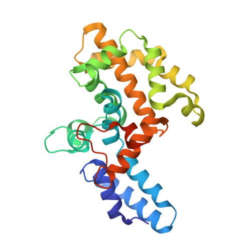
High-resolution mapping of metal ions reveals principles of surface layer assembly in Caulobacter crescentus cells.

Herdman, M.von Kugelgen, A.Kureisaite-Ciziene, D.Duman, R.El Omari, K.Garman, E.F.Kjaer, A.Kolokouris, D.Lowe, J.Wagner, A.Stansfeld, P.J.Bharat, T.A.M.

(2022) Structure 30: 215

  • DOI: https://doi.org/10.1016/j.str.2021.10.012
  • Primary Citation of Related Structures:  
    7PEO

  • PubMed Abstract: 

    Surface layers (S-layers) are proteinaceous crystalline coats that constitute the outermost component of most prokaryotic cell envelopes. In this study, we have investigated the role of metal ions in the formation of the Caulobacter crescentus S-layer using high-resolution structural and cell biology techniques, as well as molecular simulations. Utilizing optical microscopy of fluorescently tagged S-layers, we show that calcium ions facilitate S-layer lattice formation and cell-surface binding. We report all-atom molecular dynamics simulations of the S-layer lattice, revealing the importance of bound metal ions. Finally, using electron cryomicroscopy and long-wavelength X-ray diffraction experiments, we mapped the positions of metal ions in the S-layer at near-atomic resolution, supporting our insights from the cellular and simulations data. Our findings contribute to the understanding of how C. crescentus cells form a regularly arranged S-layer on their surface, with implications on fundamental S-layer biology and the synthetic biology of self-assembling biomaterials.


  • Organizational Affiliation

    Sir William Dunn School of Pathology, University of Oxford, Oxford OX1 3RE, UK.


Macromolecules
Find similar proteins by:  (by identity cutoff)  |  3D Structure
Entity ID: 1
MoleculeChains Sequence LengthOrganismDetailsImage
S-layer protein255Caulobacter vibrioidesMutation(s): 0 
Gene Names: rsaACC_1007
UniProt
Find proteins for P35828 (Caulobacter vibrioides (strain ATCC 19089 / CB15))
Explore P35828 
Go to UniProtKB:  P35828
Entity Groups  
Sequence Clusters30% Identity50% Identity70% Identity90% Identity95% Identity100% Identity
UniProt GroupP35828
Sequence Annotations
Expand
  • Reference Sequence
Oligosaccharides

Help

Entity ID: 2
MoleculeChains Length2D Diagram Glycosylation3D Interactions
4-acetamido-4,6-dideoxy-alpha-D-mannopyranose-(1-3)-4-acetamido-4,6-dideoxy-alpha-D-mannopyranose-(1-3)-beta-D-mannopyranose-(1-3)-4-acetamido-4,6-dideoxy-alpha-D-mannopyranose-(1-3)-4-acetamido-4,6-dideoxy-alpha-D-mannopyranose-(1-3)-beta-D-mannopyranose-(1-3)-4-acetamido-4,6-dideoxy-alpha-D-mannopyranose-(1-3)-4-acetamido-4,6-dideoxy-alpha-D-mannopyranose-(1-3)-beta-D-mannopyranose-(1-3)-4-acetamido-4,6-dideoxy-alpha-D-mannopyranose-(1-3)-4-acetamido-4,6-dideoxy-alpha-D-mannopyranose-(1-3)-beta-D-mannopyranose
B
12N/A
Glycosylation Resources
GlyTouCan:  G50564AO
GlyCosmos:  G50564AO
Experimental Data & Validation

Experimental Data

  • Method: ELECTRON MICROSCOPY
  • Resolution: 4.37 Å
  • Aggregation State: PARTICLE 
  • Reconstruction Method: SINGLE PARTICLE 
EM Software:
TaskSoftware PackageVersion
RECONSTRUCTIONRELION3.0
MODEL REFINEMENTREFMAC5.8.0258

Structure Validation

View Full Validation Report


Entry History & Funding Information

Deposition Data


Funding OrganizationLocationGrant Number
Wellcome TrustUnited Kingdom202231/Z/16/Z
Leverhulme TrustUnited KingdomPhilip Leverhulme Prize

Revision History  (Full details and data files)

  • Version 1.0: 2021-12-01
    Type: Initial release
  • Version 1.1: 2022-02-16
    Changes: Database references, Derived calculations