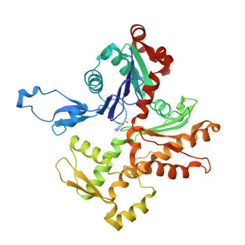
A barbed end interference mechanism reveals how capping protein promotes nucleation in branched actin networks.
Funk, J., Merino, F., Schaks, M., Rottner, K., Raunser, S., Bieling, P.(2021) Nat Commun 12: 5329-5329
- PubMed: 34504078 Search on PubMedSearch on PubMed Central
- DOI: https://doi.org/10.1038/s41467-021-25682-5
- Primary Citation Related Structures:
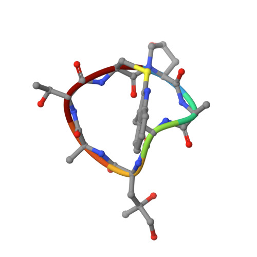
7PDZ - PubMed Abstract:
Heterodimeric capping protein (CP/CapZ) is an essential factor for the assembly of branched actin networks, which push against cellular membranes to drive a large variety of cellular processes. Aside from terminating filament growth, CP potentiates the nucleation of actin filaments by the Arp2/3 complex in branched actin networks through an unclear mechanism. Here, we combine structural biology with in vitro reconstitution to demonstrate that CP not only terminates filament elongation, but indirectly stimulates the activity of Arp2/3 activating nucleation promoting factors (NPFs) by preventing their association to filament barbed ends. Key to this function is one of CP's C-terminal "tentacle" extensions, which sterically masks the main interaction site of the terminal actin protomer. Deletion of the β tentacle only modestly impairs capping. However, in the context of a growing branched actin network, its removal potently inhibits nucleation promoting factors by tethering them to capped filament ends. End tethering of NPFs prevents their loading with actin monomers required for activation of the Arp2/3 complex and thus strongly inhibits branched network assembly both in cells and reconstituted motility assays. Our results mechanistically explain how CP couples two opposed processes-capping and nucleation-in branched actin network assembly.
- Department of Systemic Cell Biology, Max Planck Institute of Molecular Physiology, Dortmund, Germany.
Organizational Affiliation: