7PCW | pdb_00007pcw

X-RAY STRUCTURE OF THE HALOALKANE DEHALOGENASE HALOTAG7-M175W LABELED WITH A CHLOROALKANE-TETRAMETHYLRHODAMINE FLUOROPHORE SUBSTRATE


Experimental Data Snapshot

  • Method: X-RAY DIFFRACTION
  • Resolution: 2.30 Å
  • R-Value Free: 
    0.243 (Depositor), 0.241 (DCC) 
  • R-Value Work: 
    0.191 (Depositor), 0.190 (DCC) 
  • R-Value Observed: 
    0.193 (Depositor) 

Starting Model: experimental
View more details

wwPDB Validation 3D Report Full Report

Validation slider image for 7PCW

Ligand Structure Quality Assessment 


This is version 1.3 of the entry. See complete history

Literature

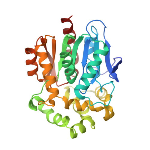
Engineered HaloTag variants for fluorescence lifetime multiplexing.

Frei, M.S.Tarnawski, M.Roberti, M.J.Koch, B.Hiblot, J.Johnsson, K.

(2022) Nat Methods 19: 65-70

  • DOI: https://doi.org/10.1038/s41592-021-01341-x
  • Primary Citation Related Structures: 
    6ZVY, 7PCW, 7PCX

  • PubMed Abstract: 

    Self-labeling protein tags such as HaloTag are powerful tools that can label fusion proteins with synthetic fluorophores for use in fluorescence microscopy. Here we introduce HaloTag variants with either increased or decreased brightness and fluorescence lifetime compared with HaloTag7 when labeled with rhodamines. Combining these HaloTag variants enabled live-cell fluorescence lifetime multiplexing of three cellular targets in one spectral channel using a single fluorophore and the generation of a fluorescence lifetime-based biosensor. Additionally, the brightest HaloTag variant showed up to 40% higher brightness in live-cell imaging applications.


  • Organizational Affiliation
    • Department of Chemical Biology, Max Planck Institute for Medical Research, Heidelberg, Germany.

Macromolecule Content 

  • Total Structure Weight: 135.81 kDa 
  • Atom Count: 9,711 
  • Modeled Residue Count: 1,170 
  • Deposited Residue Count: 1,172 
  • Unique protein chains: 1

Macromolecules

Find similar proteins by:|  3D Structure
Entity ID: 1
MoleculeChains  Sequence LengthOrganismDetailsImage
Haloalkane dehalogenase
A, B, C, D
293Rhodococcus sp. (in: high G+C Gram-positive bacteria)Mutation(s): 21 
Gene Names: dhaA
EC: 3.8.1.5
UniProt
Find proteins for P0A3G3 (Rhodococcus sp)
Explore P0A3G3 
Go to UniProtKB:  P0A3G3
Entity Groups
Sequence Clusters30% Identity50% Identity70% Identity90% Identity95% Identity100% Identity
UniProt GroupP0A3G3
Sequence Annotations
Expand
Reference Sequence

Small Molecules

Ligands 2 Unique
IDChains Name / Formula / InChI Key2D Diagram3D Interactions
OEH
(Subject of Investigation/LOI)

Query on OEH


Download:Ideal Coordinates CCD File
E [auth A],
G [auth B],
I [auth C],
K [auth D]
[9-[2-carboxy-5-[2-[2-(6-chloranylhexoxy)ethoxy]ethylcarbamoyl]phenyl]-6-(dimethylamino)xanthen-3-ylidene]-dimethyl-azanium
C35 H43 Cl N3 O6
YORHEDWUCXZZLS-UHFFFAOYSA-O
CL

Query on CL


Download:Ideal Coordinates CCD File
F [auth A],
H [auth B],
J [auth C],
L [auth D]
CHLORIDE ION
Cl
VEXZGXHMUGYJMC-UHFFFAOYSA-M

Experimental Data & Validation

Experimental Data

  • Method: X-RAY DIFFRACTION
  • Resolution: 2.30 Å
  • R-Value Free:  0.243 (Depositor), 0.241 (DCC) 
  • R-Value Work:  0.191 (Depositor), 0.190 (DCC) 
  • R-Value Observed: 0.193 (Depositor) 
Space Group: P 1
Unit Cell:
Length ( Å )Angle ( ˚ )
a = 44.32α = 68.397
b = 76.56β = 79.806
c = 85.76γ = 79.021
Software Package:
Software NamePurpose
PHENIXrefinement
XDSdata reduction
XSCALEdata scaling
PHASERphasing

Structure Validation

View Full Validation Report


Ligand Structure Quality Assessment 


Entry History 

& Funding Information

Deposition Data


Funding OrganizationLocationGrant Number
Not funded--

Revision History  (Full details and data files)

  • Version 1.0: 2021-12-15
    Type: Initial release
  • Version 1.1: 2022-02-16
    Changes: Database references
  • Version 1.2: 2024-01-31
    Changes: Data collection, Refinement description
  • Version 1.3: 2024-11-13
    Changes: Structure summary