7PBP | pdb_00007pbp

RuvAB branch migration motor complexed to the Holliday junction - RuvB AAA+ state s5 [t2 dataset]


Experimental Data Snapshot

  • Method: ELECTRON MICROSCOPY
  • Resolution: 3.20 Å
  • Aggregation State: PARTICLE 
  • Reconstruction Method: SINGLE PARTICLE 

wwPDB Validation 3D Report Full Report

Validation slider image for 7PBP

This is version 1.3 of the entry. See complete history

Literature

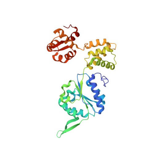
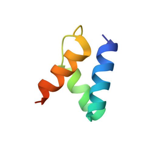
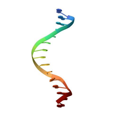
Mechanism of AAA+ ATPase-mediated RuvAB-Holliday junction branch migration.

Wald, J.Fahrenkamp, D.Goessweiner-Mohr, N.Lugmayr, W.Ciccarelli, L.Vesper, O.Marlovits, T.C.

(2022) Nature 609: 630-639

  • DOI: https://doi.org/10.1038/s41586-022-05121-1
  • Primary Citation Related Structures: 
    7PBL, 7PBM, 7PBN, 7PBO, 7PBP, 7PBQ, 7PBR, 7PBS, 7PBT, 7PBU

  • PubMed Abstract: 

    The Holliday junction is a key intermediate formed during DNA recombination across all kingdoms of life 1 . In bacteria, the Holliday junction is processed by two homo-hexameric AAA+ ATPase RuvB motors, which assemble together with the RuvA-Holliday junction complex to energize the strand-exchange reaction 2 . Despite its importance for chromosome maintenance, the structure and mechanism by which this complex facilitates branch migration are unknown. Here, using time-resolved cryo-electron microscopy, we obtained structures of the ATP-hydrolysing RuvAB complex in seven distinct conformational states, captured during assembly and processing of a Holliday junction. Five structures together resolve the complete nucleotide cycle and reveal the spatiotemporal relationship between ATP hydrolysis, nucleotide exchange and context-specific conformational changes in RuvB. Coordinated motions in a converter formed by DNA-disengaged RuvB subunits stimulate hydrolysis and nucleotide exchange. Immobilization of the converter enables RuvB to convert the ATP-contained energy into a lever motion, which generates the pulling force driving the branch migration. We show that RuvB motors rotate together with the DNA substrate, which, together with a progressing nucleotide cycle, forms the mechanistic basis for DNA recombination by continuous branch migration. Together, our data decipher the molecular principles of homologous recombination by the RuvAB complex, elucidate discrete and sequential transition-state intermediates for chemo-mechanical coupling of hexameric AAA+ motors and provide a blueprint for the design of state-specific compounds targeting AAA+ motors.


  • Organizational Affiliation
    • Institute of Structural and Systems Biology, University Medical Center Hamburg-Eppendorf, Hamburg, Germany. jiri.wald@cssb-hamburg.de.

Macromolecule Content 

  • Total Structure Weight: 236.66 kDa 
  • Atom Count: 16,259 
  • Modeled Residue Count: 1,998 
  • Deposited Residue Count: 2,028 
  • Unique protein chains: 2
  • Unique nucleic acid chains: 2

Macromolecules


Find similar proteins by:|  3D Structure
Entity ID: 1
MoleculeChains  Sequence LengthOrganismDetailsImage
Holliday junction ATP-dependent DNA helicase RuvB
A, B, C, D, E
A, B, C, D, E, F
315Streptococcus thermophilusMutation(s): 0 
Gene Names: ruvBCDA68_01670STHERMO_2112
EC: 3.6.4.12 (PDB Primary Data), 3.6.4 (UniProt)
UniProt
Find proteins for Q5M2B1 (Streptococcus thermophilus (strain ATCC BAA-250 / LMG 18311))
Explore Q5M2B1 
Go to UniProtKB:  Q5M2B1
Entity Groups
Sequence Clusters30% Identity50% Identity70% Identity90% Identity95% Identity100% Identity
UniProt GroupQ5M2B1
Sequence Annotations
Expand
Reference Sequence
Find similar proteins by:|  3D Structure
Entity ID: 2
MoleculeChains  Sequence LengthOrganismDetailsImage
Holliday junction ATP-dependent DNA helicase RuvA
G, H
54Salmonella enterica subsp. enterica serovar TyphimuriumMutation(s): 0 
Gene Names: ruvA
EC: 3.6.4.12
UniProt
Find proteins for P66746 (Salmonella typhimurium (strain LT2 / SGSC1412 / ATCC 700720))
Explore P66746 
Go to UniProtKB:  P66746
Entity Groups
Sequence Clusters30% Identity50% Identity70% Identity90% Identity95% Identity100% Identity
UniProt GroupP66746
Sequence Annotations
Expand
Reference Sequence
Find similar nucleic acids by:  Sequence
Entity ID: 3
MoleculeChains LengthOrganismImage
random DNAI [auth U]15synthetic construct
Sequence Annotations
Expand
Reference Sequence
Find similar nucleic acids by:  Sequence
Entity ID: 4
MoleculeChains LengthOrganismImage
random DNAJ [auth V]15synthetic construct
Sequence Annotations
Expand
Reference Sequence

Small Molecules

Ligands 3 Unique
IDChains Name / Formula / InChI Key2D Diagram3D Interactions
AGS

Query on AGS


Download:Ideal Coordinates CCD File
L [auth B],
N [auth C],
P [auth D]
PHOSPHOTHIOPHOSPHORIC ACID-ADENYLATE ESTER
C10 H16 N5 O12 P3 S
NLTUCYMLOPLUHL-KQYNXXCUSA-N
ADP

Query on ADP


Download:Ideal Coordinates CCD File
K [auth A],
R [auth E],
S [auth F]
ADENOSINE-5'-DIPHOSPHATE
C10 H15 N5 O10 P2
XTWYTFMLZFPYCI-KQYNXXCUSA-N
MG

Query on MG


Download:Ideal Coordinates CCD File
M [auth B],
O [auth C],
Q [auth D]
MAGNESIUM ION
Mg
JLVVSXFLKOJNIY-UHFFFAOYSA-N

Experimental Data & Validation

Experimental Data

  • Method: ELECTRON MICROSCOPY
  • Resolution: 3.20 Å
  • Aggregation State: PARTICLE 
  • Reconstruction Method: SINGLE PARTICLE 
EM Software:
TaskSoftware PackageVersion
MODEL REFINEMENTPHENIX
RECONSTRUCTIONRELION3

Structure Validation

View Full Validation Report


Entry History 

Deposition Data

Revision History  (Full details and data files)

  • Version 1.0: 2022-09-14
    Type: Initial release
  • Version 1.1: 2022-09-28
    Changes: Database references
  • Version 1.2: 2022-11-30
    Changes: Database references
  • Version 1.3: 2024-07-17
    Changes: Data collection