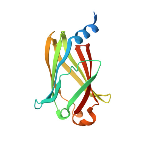
7PAC

The crystal structure of PDE6D in the apo state


Experimental Data Snapshot

  • Method: X-RAY DIFFRACTION
  • Resolution: 2.05 Å
  • R-Value Free: 0.232 
  • R-Value Work: 0.185 
  • R-Value Observed: 0.188 

Starting Model: experimental
View more details

wwPDB Validation   3D Report Full Report


Ligand Structure Quality Assessment 


This is version 1.1 of the entry. See complete history


Literature

Validation of a small molecule inhibitor of PDE6D-RAS interaction with favorable anti-leukemic effects.

Canovas Nunes, S.De Vita, S.Anighoro, A.Autelitano, F.Beaumont, E.Klingbeil, P.McGuinness, M.Duvert, B.Harris, C.Yang, L.Pokharel, S.P.Chen, C.W.Ermann, M.Williams, D.A.Xu, H.

(2022) Blood Cancer J 12: 64-64

  • DOI: https://doi.org/10.1038/s41408-022-00663-z
  • Primary Citation of Related Structures:  
    7PAC, 7PAD, 7PAE

  • PubMed Abstract: 

    RAS mutations prevalent in high-risk leukemia have been linked to relapse and chemotherapy resistance. Efforts to directly target RAS proteins have been largely unsuccessful. However, since RAS-mediated transformation is dependent on signaling through the RAS-related C3 botulinum toxin substrate (RAC) small GTPase, we hypothesized that targeting RAC may be an effective therapeutic approach in RAS mutated tumors. Here we describe multiple small molecules capable of inhibiting RAC activation in acute lymphoblastic leukemia cell lines. One of these, DW0254, also demonstrates promising anti-leukemic activity in RAS-mutated cells. Using chemical proteomics and biophysical methods, we identified the hydrophobic pocket of phosphodiester 6 subunit delta (PDE6D), a known RAS chaperone, as a target for this compound. Inhibition of RAS localization to the plasma membrane upon DW0254 treatment is associated with RAC inhibition through a phosphatidylinositol-3-kinase/AKT-dependent mechanism. Our findings provide new insights into the importance of PDE6D-mediated transport for RAS-dependent RAC activation and leukemic cell survival.


  • Organizational Affiliation

    Division of Hematology/Oncology, Boston Children's Hospital, Harvard Medical School, Boston, MA, USA.


Macromolecules
Find similar proteins by:  (by identity cutoff)  |  3D Structure
Entity ID: 1
MoleculeChains Sequence LengthOrganismDetailsImage
Retinal rod rhodopsin-sensitive cGMP 3',5'-cyclic phosphodiesterase subunit deltaA [auth B]166Homo sapiensMutation(s): 0 
Gene Names: PDE6DPDED
UniProt & NIH Common Fund Data Resources
Find proteins for O43924 (Homo sapiens)
Explore O43924 
Go to UniProtKB:  O43924
PHAROS:  O43924
GTEx:  ENSG00000156973 
Entity Groups  
Sequence Clusters30% Identity50% Identity70% Identity90% Identity95% Identity100% Identity
UniProt GroupO43924
Sequence Annotations
Expand
  • Reference Sequence
Experimental Data & Validation

Experimental Data

  • Method: X-RAY DIFFRACTION
  • Resolution: 2.05 Å
  • R-Value Free: 0.232 
  • R-Value Work: 0.185 
  • R-Value Observed: 0.188 
  • Space Group: P 1 21 1
Unit Cell:
Length ( Å )Angle ( ˚ )
a = 44.25α = 90
b = 39.18β = 105.89
c = 44.68γ = 90
Software Package:
Software NamePurpose
REFMACrefinement
PDB_EXTRACTdata extraction
xia2data reduction
Aimlessdata scaling
DIMPLEphasing

Structure Validation

View Full Validation Report


Ligand Structure Quality Assessment 


Entry History & Funding Information

Deposition Data


Funding OrganizationLocationGrant Number
National Institutes of Health/National Cancer Institute (NIH/NCI)5R01CA202756

Revision History  (Full details and data files)

  • Version 1.0: 2022-06-08
    Type: Initial release
  • Version 1.1: 2024-01-31
    Changes: Data collection, Refinement description