7P4T | pdb_00007p4t

Tetrameric structure of murine SapA


Experimental Data Snapshot

  • Method: X-RAY DIFFRACTION
  • Resolution: 3.17 Å
  • R-Value Free: 
    0.304 (Depositor) 
  • R-Value Work: 
    0.272 (Depositor), 0.276 (DCC) 

Starting Model: experimental
View more details

wwPDB Validation 3D Report Full Report

Validation slider image for 7P4T

This is version 1.3 of the entry. See complete history

Literature

A Tetrameric Assembly of Saposin A: Increasing Structural Diversity in Lipid Transfer Proteins.

Shamin, M.Spratley, S.J.Graham, S.C.Deane, J.E.

(2021) Contact (Thousand Oaks) 4: 251525642110523-251525642110523

  • DOI: https://doi.org/10.1177/25152564211052382
  • Primary Citation Related Structures: 
    7P4T

  • PubMed Abstract: 

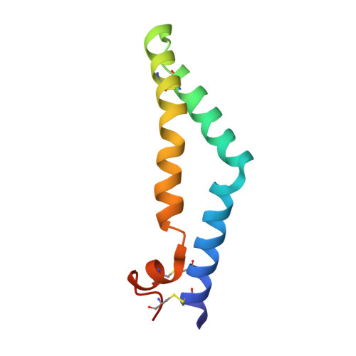
    Saposins are lipid transfer proteins required for the degradation of sphingolipids in the lysosome. These small proteins bind lipids by transitioning from a closed, monomeric state to an open conformation exposing a hydrophobic surface that binds and shields hydrophobic lipid tails from the aqueous environment. Saposins form a range of multimeric assemblies to encompass these bound lipids and present them to hydrolases in the lysosome. This lipid-binding property of human saposin A has been exploited to form lipoprotein nanodiscs suitable for structural studies of membrane proteins. Here we present the crystal structure of a unique tetrameric assembly of murine saposin A produced serendipitously, following modifications of published protocols for making lipoprotein nanodiscs. The structure of this new saposin oligomer highlights the diversity of tertiary arrangement that can be adopted by these important lipid transfer proteins.


  • Organizational Affiliation
    • Cambridge Institute for Medical Research, University of Cambridge, Cambridge CB2 0XY, UK.

Macromolecule Content 

  • Total Structure Weight: 36.49 kDa 
  • Atom Count: 2,432 
  • Modeled Residue Count: 320 
  • Deposited Residue Count: 332 
  • Unique protein chains: 1

Macromolecules

Find similar proteins by:|  3D Structure
Entity ID: 1
MoleculeChains  Sequence LengthOrganismDetailsImage
Saposin-A
A, B, C, D
83Mus musculusMutation(s): 0 
Gene Names: PsapSgp1
UniProt
Find proteins for Q61207 (Mus musculus)
Explore Q61207 
Go to UniProtKB:  Q61207
Entity Groups
Sequence Clusters30% Identity50% Identity70% Identity90% Identity95% Identity100% Identity
UniProt GroupQ61207
Sequence Annotations
Expand
Reference Sequence

Experimental Data & Validation

Experimental Data

  • Method: X-RAY DIFFRACTION
  • Resolution: 3.17 Å
  • R-Value Free:  0.304 (Depositor) 
  • R-Value Work:  0.272 (Depositor), 0.276 (DCC) 
Space Group: P 65
Unit Cell:
Length ( Å )Angle ( ˚ )
a = 48.489α = 90
b = 48.489β = 90
c = 314.359γ = 120
Software Package:
Software NamePurpose
DIALSdata collection
autoPROCdata scaling
PHASERphasing
PHENIXrefinement

Structure Validation

View Full Validation Report


Entry History 

& Funding Information

Deposition Data


Funding OrganizationLocationGrant Number
Wellcome TrustUnited Kingdom--

Revision History  (Full details and data files)

  • Version 1.0: 2022-05-25
    Type: Initial release
  • Version 1.1: 2023-05-17
    Changes: Database references
  • Version 1.2: 2024-02-07
    Changes: Data collection, Refinement description
  • Version 1.3: 2024-10-16
    Changes: Structure summary