7P4J | pdb_00007p4j

Crystal structure of Autotaxin and tetrahydrocannabinol


Experimental Data Snapshot

  • Method: X-RAY DIFFRACTION
  • Resolution: 1.79 Å
  • R-Value Free: 
    0.241 (Depositor), 0.241 (DCC) 
  • R-Value Work: 
    0.183 (Depositor), 0.183 (DCC) 
  • R-Value Observed: 
    0.185 (Depositor) 

Starting Model: experimental
View more details

wwPDB Validation 3D Report Full Report

Validation slider image for 7P4J

Ligand Structure Quality Assessment 


This is version 1.3 of the entry. See complete history

Literature

Linking medicinal cannabis to autotaxin-lysophosphatidic acid signaling.

Eymery, M.C.McCarthy, A.A.Hausmann, J.

(2023) Life Sci Alliance 6

  • DOI: https://doi.org/10.26508/lsa.202201595
  • Primary Citation Related Structures: 
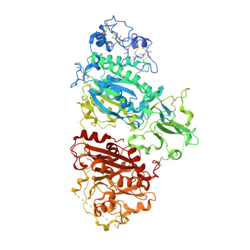
    7P4J, 7P4O

  • PubMed Abstract: 

    Autotaxin is primarily known for the formation of lysophosphatidic acid (LPA) from lysophosphatidylcholine. LPA is an important signaling phospholipid that can bind to six G protein-coupled receptors (LPA 1-6 ). The ATX-LPA signaling axis is a critical component in many physiological and pathophysiological conditions. Here, we describe a potent inhibition of Δ 9 - trans -tetrahydrocannabinol (THC), the main psychoactive compound of medicinal cannabis and related cannabinoids, on the catalysis of two isoforms of ATX with nanomolar apparent EC 50 values. Furthermore, we decipher the binding interface of ATX to THC, and its derivative 9(R)-Δ6a,10a-THC (6a10aTHC), by X-ray crystallography. Cellular experiments confirm this inhibitory effect, revealing a significant reduction of internalized LPA 1 in the presence of THC with simultaneous ATX and lysophosphatidylcholine stimulation. Our results establish a functional interaction of THC with autotaxin-LPA signaling and highlight novel aspects of medicinal cannabis therapy.


  • Organizational Affiliation
    • European Molecular Biology Laboratory, Grenoble, Grenoble, France.

Macromolecule Content 

  • Total Structure Weight: 99.77 kDa 
  • Atom Count: 6,792 
  • Modeled Residue Count: 770 
  • Deposited Residue Count: 827 
  • Unique protein chains: 1

Macromolecules

Find similar proteins by:|  3D Structure
Entity ID: 1
MoleculeChains  Sequence LengthOrganismDetailsImage
Ectonucleotide pyrophosphatase/phosphodiesterase family member 2827Rattus norvegicusMutation(s): 4 
Gene Names: Enpp2AtxNpps2
EC: 3.1.4.39 (PDB Primary Data), 3.1.4.4 (UniProt)
UniProt
Find proteins for Q64610 (Rattus norvegicus)
Explore Q64610 
Go to UniProtKB:  Q64610
Entity Groups
Sequence Clusters30% Identity50% Identity70% Identity90% Identity95% Identity100% Identity
UniProt GroupQ64610
Glycosylation
Glycosylation Sites: 1Go to GlyGen: Q64610-1
Sequence Annotations
Expand
Reference Sequence

Oligosaccharides

Help  
Entity ID: 2
MoleculeChains Length2D Diagram GlycosylationD Interactions
2-acetamido-2-deoxy-beta-D-glucopyranose-(1-4)-2-acetamido-2-deoxy-beta-D-glucopyranose
B
2N-Glycosylation
Glycosylation Resources
GlyTouCan: G42666HT
GlyCosmos: G42666HT
GlyGen: G42666HT

Small Molecules

Ligands 9 Unique
IDChains Name / Formula / InChI Key2D Diagram3D Interactions
5JK

Query on 5JK


Download:Ideal Coordinates CCD File
F [auth A]7alpha-hydroxycholesterol
C27 H46 O2
OYXZMSRRJOYLLO-RVOWOUOISA-N
TCI
(Subject of Investigation/LOI)

Query on TCI


Download:Ideal Coordinates CCD File
E [auth A](6aR,10aR)-6,6,9-trimethyl-3-pentyl-6a,7,8,10a-tetrahydro-6H-benzo[c]chromen-1-ol
C21 H30 O2
CYQFCXCEBYINGO-IAGOWNOFSA-N
IOD

Query on IOD


Download:Ideal Coordinates CCD File
AA [auth A]
BA [auth A]
CA [auth A]
DA [auth A]
EA [auth A]
AA [auth A],
BA [auth A],
CA [auth A],
DA [auth A],
EA [auth A],
FA [auth A],
G [auth A],
GA [auth A],
H [auth A],
HA [auth A],
I [auth A],
IA [auth A],
J [auth A],
JA [auth A],
V [auth A],
W [auth A],
X [auth A],
Y [auth A],
Z [auth A]
IODIDE ION
I
XMBWDFGMSWQBCA-UHFFFAOYSA-M
PO4

Query on PO4


Download:Ideal Coordinates CCD File
Q [auth A]PHOSPHATE ION
O4 P
NBIIXXVUZAFLBC-UHFFFAOYSA-K
GOL

Query on GOL


Download:Ideal Coordinates CCD File
L [auth A]
M [auth A]
N [auth A]
O [auth A]
P [auth A]
L [auth A],
M [auth A],
N [auth A],
O [auth A],
P [auth A],
R [auth A],
T [auth A],
U [auth A]
GLYCEROL
C3 H8 O3
PEDCQBHIVMGVHV-UHFFFAOYSA-N
ZN

Query on ZN


Download:Ideal Coordinates CCD File
C [auth A],
D [auth A]
ZINC ION
Zn
PTFCDOFLOPIGGS-UHFFFAOYSA-N
SCN

Query on SCN


Download:Ideal Coordinates CCD File
K [auth A],
S [auth A]
THIOCYANATE ION
C N S
ZMZDMBWJUHKJPS-UHFFFAOYSA-M
CA

Query on CA


Download:Ideal Coordinates CCD File
KA [auth A],
LA [auth A]
CALCIUM ION
Ca
BHPQYMZQTOCNFJ-UHFFFAOYSA-N
NA

Query on NA


Download:Ideal Coordinates CCD File
MA [auth A]SODIUM ION
Na
FKNQFGJONOIPTF-UHFFFAOYSA-N
Binding Affinity Annotations 
IDSourceBinding Affinity
TCI BindingDB:  7P4J EC50: 460 (nM) from 1 assay(s)

Experimental Data & Validation

Experimental Data

  • Method: X-RAY DIFFRACTION
  • Resolution: 1.79 Å
  • R-Value Free:  0.241 (Depositor), 0.241 (DCC) 
  • R-Value Work:  0.183 (Depositor), 0.183 (DCC) 
  • R-Value Observed: 0.185 (Depositor) 
Space Group: P 1
Unit Cell:
Length ( Å )Angle ( ˚ )
a = 53.705α = 103.154
b = 60.979β = 97.394
c = 63.603γ = 94.228
Software Package:
Software NamePurpose
PHENIXrefinement
MxCuBEdata collection
autoPROCdata processing
Cootmodel building
XDSdata reduction
Aimlessdata scaling
pointlessdata scaling
PHENIXphasing
STARANISOdata scaling

Structure Validation

View Full Validation Report


Ligand Structure Quality Assessment 


Entry History 

& Funding Information

Deposition Data


Funding OrganizationLocationGrant Number
EIPOD fellowship under Marie Sklodowska-Curie Actions COFUNDEuropean Union664726

Revision History  (Full details and data files)

  • Version 1.0: 2022-12-28
    Type: Initial release
  • Version 1.1: 2023-01-25
    Changes: Database references
  • Version 1.2: 2024-02-07
    Changes: Data collection, Refinement description
  • Version 1.3: 2024-10-23
    Changes: Structure summary