7P46 | pdb_00007p46

Crystal Structure of Xanthomonas campestris Tryptophan 2,3-dioxygenase (TDO)


Experimental Data Snapshot

  • Method: X-RAY DIFFRACTION
  • Resolution: 1.70 Å
  • R-Value Free: 
    0.185 (Depositor), 0.195 (DCC) 
  • R-Value Work: 
    0.149 (Depositor), 0.162 (DCC) 
  • R-Value Observed: 
    0.151 (Depositor) 

Starting Model: experimental
View more details

wwPDB Validation 3D Report Full Report

Validation slider image for 7P46

Ligand Structure Quality Assessment 


This is version 1.1 of the entry. See complete history

Literature

Binding of l-kynurenine to X. campestris tryptophan 2,3-dioxygenase.

Basran, J.Booth, E.S.Campbell, L.P.Thackray, S.J.Jesani, M.H.Clayden, J.Moody, P.C.E.Mowat, C.G.Kwon, H.Raven, E.L.

(2021) J Inorg Biochem 225: 111604-111604

  • DOI: https://doi.org/10.1016/j.jinorgbio.2021.111604
  • Primary Citation Related Structures: 
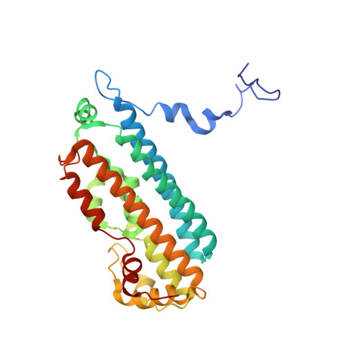
    7P46

  • PubMed Abstract: 

    The kynurenine pathway is the major route of tryptophan metabolism. The first step of this pathway is catalysed by one of two heme-dependent dioxygenase enzymes - tryptophan 2,3-dioxygenase (TDO) and indoleamine 2,3-dioxygenase (IDO) - leading initially to the formation of N-formylkynurenine (NFK). In this paper, we present a crystal structure of a bacterial TDO from X. campestris in complex with l-kynurenine, the hydrolysed product of NFK. l-kynurenine is bound at the active site in a similar location to the substrate (l-Trp). Hydrogen bonding interactions with Arg117 and the heme 7-propionate anchor the l-kynurenine molecule into the pocket. A mechanism for the hydrolysis of NFK in the active site is presented.


  • Organizational Affiliation
    • Department of Molecular and Cell Biology, Leicester Institute of Structural and Chemical Biology, University of Leicester, Leicester LE1 9HN, UK.

Macromolecule Content 

  • Total Structure Weight: 273.22 kDa 
  • Atom Count: 21,574 
  • Modeled Residue Count: 2,235 
  • Deposited Residue Count: 2,256 
  • Unique protein chains: 1

Macromolecules

Find similar proteins by:|  3D Structure
Entity ID: 1
MoleculeChains  Sequence LengthOrganismDetailsImage
Tryptophan 2,3-dioxygenase
A, B, C, D, E
A, B, C, D, E, F, G, H
282Xanthomonas campestris pv. campestris str. ATCC 33913Mutation(s): 0 
Gene Names: kynAXCC0432
EC: 1.13.11.11
UniProt
Find proteins for Q8PDA8 (Xanthomonas campestris pv. campestris (strain ATCC 33913 / DSM 3586 / NCPPB 528 / LMG 568 / P 25))
Explore Q8PDA8 
Go to UniProtKB:  Q8PDA8
Entity Groups
Sequence Clusters30% Identity50% Identity70% Identity90% Identity95% Identity100% Identity
UniProt GroupQ8PDA8
Sequence Annotations
Expand
Reference Sequence

Small Molecules

Ligands 5 Unique
IDChains Name / Formula / InChI Key2D Diagram3D Interactions
HEM
(Subject of Investigation/LOI)

Query on HEM


Download:Ideal Coordinates CCD File
EA [auth D]
I [auth A]
IA [auth E]
LA [auth F]
QA [auth G]
EA [auth D],
I [auth A],
IA [auth E],
LA [auth F],
QA [auth G],
S [auth B],
TA [auth H],
Y [auth C]
PROTOPORPHYRIN IX CONTAINING FE
C34 H32 Fe N4 O4
KABFMIBPWCXCRK-RGGAHWMASA-L
KYN

Query on KYN


Download:Ideal Coordinates CCD File
AA [auth C]
GA [auth D]
JA [auth E]
K [auth A]
NA [auth F]
AA [auth C],
GA [auth D],
JA [auth E],
K [auth A],
NA [auth F],
RA [auth G],
U [auth B],
VA [auth H]
(2S)-2-amino-4-(2-aminophenyl)-4-oxobutanoic acid
C10 H12 N2 O3
YGPSJZOEDVAXAB-QMMMGPOBSA-N
TRP

Query on TRP


Download:Ideal Coordinates CCD File
BA [auth C]
HA [auth D]
KA [auth E]
L [auth A]
OA [auth F]
BA [auth C],
HA [auth D],
KA [auth E],
L [auth A],
OA [auth F],
SA [auth G],
V [auth B],
WA [auth H]
TRYPTOPHAN
C11 H12 N2 O2
QIVBCDIJIAJPQS-VIFPVBQESA-N
GOL

Query on GOL


Download:Ideal Coordinates CCD File
CA [auth C]
DA [auth C]
M [auth A]
N [auth A]
O [auth A]
CA [auth C],
DA [auth C],
M [auth A],
N [auth A],
O [auth A],
P [auth A],
PA [auth F],
Q [auth A],
R [auth A],
W [auth B],
X [auth B],
XA [auth H],
YA [auth H],
ZA [auth H]
GLYCEROL
C3 H8 O3
PEDCQBHIVMGVHV-UHFFFAOYSA-N
CYN

Query on CYN


Download:Ideal Coordinates CCD File
FA [auth D]
J [auth A]
MA [auth F]
T [auth B]
UA [auth H]
FA [auth D],
J [auth A],
MA [auth F],
T [auth B],
UA [auth H],
Z [auth C]
CYANIDE ION
C N
XFXPMWWXUTWYJX-UHFFFAOYSA-N

Experimental Data & Validation

Experimental Data

  • Method: X-RAY DIFFRACTION
  • Resolution: 1.70 Å
  • R-Value Free:  0.185 (Depositor), 0.195 (DCC) 
  • R-Value Work:  0.149 (Depositor), 0.162 (DCC) 
  • R-Value Observed: 0.151 (Depositor) 
Space Group: P 1 21 1
Unit Cell:
Length ( Å )Angle ( ˚ )
a = 78.095α = 90
b = 117.752β = 95.52
c = 138.819γ = 90
Software Package:
Software NamePurpose
REFMACrefinement
PDB_EXTRACTdata extraction
XDSdata reduction
Aimlessdata scaling
PHASERphasing

Structure Validation

View Full Validation Report


Ligand Structure Quality Assessment 


Entry History 

Deposition Data

Revision History  (Full details and data files)

  • Version 1.0: 2021-10-06
    Type: Initial release
  • Version 1.1: 2024-01-31
    Changes: Data collection, Refinement description