Structure-Guided Design of d-Galactal Derivatives with High Affinity and Selectivity for the Galectin-8 N-Terminal Domain.
Hassan, M., Baussiere, F., Guzelj, S., Sundin, A.P., Hakansson, M., Kovacic, R., Leffler, H., Tomasic, T., Anderluh, M., Jakopin, Z., Nilsson, U.J.(2021) ACS Med Chem Lett 12: 1745-1752
- PubMed: 34795863 Search on PubMedSearch on PubMed Central
- DOI: https://doi.org/10.1021/acsmedchemlett.1c00371
- Primary Citation Related Structures:
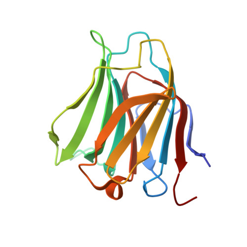
7P11, 7P1M - PubMed Abstract:
Galectin-8 is a carbohydrate-binding protein that plays a crucial role in tumor progression and metastasis, antibacterial autophagy, modulation of the immune system, and bone remodeling. The design, synthesis, and protein affinity evaluation of a set of C-3 substituted benzimidazole and quinoline d-galactal derivatives identified a d-galactal-benzimidazole hybrid as a selective ligand for the galectin-8 N-terminal domain (galectin-8N), with a K d of 48 μM and 15-fold selectivity over galectin-3 and even better selectivity over the other mammalian galectins. X-ray structural analysis of galectin-8N in complex with one benzimidazole- and one quinoline-galactal derivative at 1.52 and 2.1 Å together with molecular dynamics simulations and quantum mechanical calculations of galectin-8N in complex with the benzimidazole derivative revealed orbital overlap between a NH LUMO of Arg45 with electron rich HOMOs of the olefin and O4 of the d-galactal. Such overlap is hypothesized to contribute to the high affinity of the d-galactal-derived ligands for galectin-8N. A (3-(4,5-dimethylthiazol-2-yl)-5-(3- carboxymethoxyphenyl)-2-(4-sulfophenyl)-2 H -tetrazolium) (MTS) assay evaluation of the d-galactal-benzimidazole hybrid and an analogous galactoside derivative on a panel of cell lines with MTS assay showed no effect on cell viability up to 100 μM concentration. A subsequent functional assay using the MDA-MB-231 cell line demonstrated that the d-galactal-benzimidazole hybrid and the analogous galactoside derivative reduced the secretion of the proinflammatory cytokines interleukin-6 (IL-6) and IL-8 in a dose-dependent manner. Therefore, these compounds represent potential probes for galectin-8N pharmacology investigations and possibly promising leads for the design and synthesis of potent and selective galectin-8 inhibitors as potential antitumor and anti-inflammatory agents.
- Centre for Analysis and Synthesis, Department of Chemistry, Lund University, Box 124, SE-221 00 Lund, Sweden.
Organizational Affiliation: