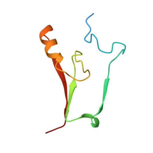
7P0O

mitoNEET bound to M1 molecule


Experimental Data Snapshot

  • Method: X-RAY DIFFRACTION
  • Resolution: 1.65 Å
  • R-Value Free: 0.236 
  • R-Value Work: 0.171 
  • R-Value Observed: 0.174 

Starting Model: experimental
View more details

wwPDB Validation   3D Report Full Report


Ligand Structure Quality Assessment 


This is version 1.1 of the entry. See complete history


Literature

An anti-diabetic drug targets NEET (CISD) proteins through destabilization of their [2Fe-2S] clusters.

Marjault, H.B.Karmi, O.Zuo, K.Michaeli, D.Eisenberg-Domovich, Y.Rossetti, G.de Chassey, B.Vonderscher, J.Cabantchik, I.Carloni, P.Mittler, R.Livnah, O.Meldrum, E.Nechushtai, R.

(2022) Commun Biol 5: 437-437

  • DOI: https://doi.org/10.1038/s42003-022-03393-x
  • Primary Citation of Related Structures:  
    7P0O, 7P0P

  • PubMed Abstract: 

    Elevated levels of mitochondrial iron and reactive oxygen species (ROS) accompany the progression of diabetes, negatively impacting insulin production and secretion from pancreatic cells. In search for a tool to reduce mitochondrial iron and ROS levels, we arrived at a molecule that destabilizes the [2Fe-2S] clusters of NEET proteins (M1). Treatment of db/db diabetic mice with M1 improved hyperglycemia, without the weight gain observed with alternative treatments such as rosiglitazone. The molecular interactions of M1 with the NEET proteins mNT and NAF-1 were determined by X-crystallography. The possibility of controlling diabetes by molecules that destabilize the [2Fe-2S] clusters of NEET proteins, thereby reducing iron-mediated oxidative stress, opens a new route for managing metabolic aberration such as in diabetes.


  • Organizational Affiliation

    The Alexander Silberman Institute of Life Science and The Wolfson Centre for Applied Structural Biology, Faculty of Science and Mathematics, The Edmond J. Safra Campus at Givat Ram, The Hebrew University of Jerusalem, Jerusalem, 91904, Israel.


Macromolecules
Find similar proteins by:  (by identity cutoff)  |  3D Structure
Entity ID: 1
MoleculeChains Sequence LengthOrganismDetailsImage
CDGSH iron-sulfur domain-containing protein 1
A, B
76Homo sapiensMutation(s): 0 
Gene Names: CISD1C10orf70ZCD1MDS029
EC: 2.6.1.3
UniProt & NIH Common Fund Data Resources
Find proteins for Q9NZ45 (Homo sapiens)
Explore Q9NZ45 
Go to UniProtKB:  Q9NZ45
PHAROS:  Q9NZ45
GTEx:  ENSG00000122873 
Entity Groups  
Sequence Clusters30% Identity50% Identity70% Identity90% Identity95% Identity100% Identity
UniProt GroupQ9NZ45
Sequence Annotations
Expand
  • Reference Sequence
Experimental Data & Validation

Experimental Data

  • Method: X-RAY DIFFRACTION
  • Resolution: 1.65 Å
  • R-Value Free: 0.236 
  • R-Value Work: 0.171 
  • R-Value Observed: 0.174 
  • Space Group: P 21 21 21
Unit Cell:
Length ( Å )Angle ( ˚ )
a = 45.152α = 90
b = 49.962β = 90
c = 58.945γ = 90
Software Package:
Software NamePurpose
REFMACrefinement
MOSFLMdata reduction
XDSdata scaling
MOLREPphasing

Structure Validation

View Full Validation Report


Ligand Structure Quality Assessment 


Entry History & Funding Information

Deposition Data


Funding OrganizationLocationGrant Number
Marie Sklodowska-Curie Actions, FragNET ITNIsrael765048

Revision History  (Full details and data files)

  • Version 1.0: 2022-05-25
    Type: Initial release
  • Version 1.1: 2024-01-31
    Changes: Data collection, Refinement description