7P0H | pdb_00007p0h

Crystal structure of Helicobacter pylori ComF fused to an artificial alphaREP crystallization helper(named B2)


Experimental Data Snapshot

  • Method: X-RAY DIFFRACTION
  • Resolution: 2.50 Å
  • R-Value Free: 
    0.245 (Depositor), 0.257 (DCC) 
  • R-Value Work: 
    0.221 (Depositor), 0.229 (DCC) 
  • R-Value Observed: 
    0.222 (Depositor) 

wwPDB Validation 3D Report Full Report

Validation slider image for 7P0H

Ligand Structure Quality Assessment 


This is version 1.2 of the entry. See complete history

Literature

ComFC mediates transport and handling of single-stranded DNA during natural transformation.

Damke, P.P.Celma, L.Kondekar, S.M.Di Guilmi, A.M.Marsin, S.Depagne, J.Veaute, X.Legrand, P.Walbott, H.Vercruyssen, J.Guerois, R.Quevillon-Cheruel, S.Radicella, J.P.

(2022) Nat Commun 13: 1961-1961

  • DOI: https://doi.org/10.1038/s41467-022-29494-z
  • Primary Citation Related Structures: 
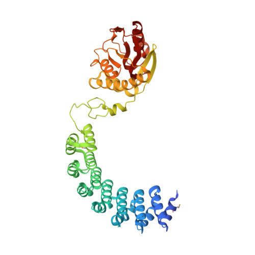
    7P0H

  • PubMed Abstract: 

    The ComFC protein is essential for natural transformation, a process that plays a major role in the spread of antibiotic resistance genes and virulence factors across bacteria. However, its role remains largely unknown. Here, we show that Helicobacter pylori ComFC is involved in DNA transport through the cell membrane, and is required for the handling of the single-stranded DNA once it is delivered into the cytoplasm. The crystal structure of ComFC includes a zinc-finger motif and a putative phosphoribosyl transferase domain, both necessary for the protein's in vivo activity. Furthermore, we show that ComFC is a membrane-associated protein with affinity for single-stranded DNA. Our results suggest that ComFC provides the link between the transport of the transforming DNA into the cytoplasm and its handling by the recombination machinery.


  • Organizational Affiliation
    • Université Paris-Saclay, CEA, Stabilité Génétique Cellules Souches et Radiations, Institut de Biologie François Jacob, F-92260, Fontenay aux Roses, France.

Macromolecule Content 

  • Total Structure Weight: 191.79 kDa 
  • Atom Count: 13,138 
  • Modeled Residue Count: 1,644 
  • Deposited Residue Count: 1,708 
  • Unique protein chains: 1

Macromolecules

Find similar proteins by:|  3D Structure
Entity ID: 1
MoleculeChains  Sequence LengthOrganismDetailsImage
Helicobacter pylori ComF fused to an artificial alphaREP crystallization helper (named B2)
A, B, C, D
427synthetic constructHelicobacter pylori 26695
This entity is chimeric
Mutation(s): 0 
UniProt
Find proteins for O26008 (Helicobacter pylori (strain ATCC 700392 / 26695))
Explore O26008 
Go to UniProtKB:  O26008
Entity Groups
Sequence Clusters30% Identity50% Identity70% Identity90% Identity95% Identity100% Identity
UniProt GroupO26008
Sequence Annotations
Expand
Reference Sequence

Small Molecules

Ligands 4 Unique
IDChains Name / Formula / InChI Key2D Diagram3D Interactions
PRP
(Subject of Investigation/LOI)

Query on PRP


Download:Ideal Coordinates CCD File
DA [auth D],
F [auth A],
P [auth B],
V [auth C]
1-O-pyrophosphono-5-O-phosphono-alpha-D-ribofuranose
C5 H13 O14 P3
PQGCEDQWHSBAJP-TXICZTDVSA-N
GOL

Query on GOL


Download:Ideal Coordinates CCD File
AA [auth C]
FA [auth D]
GA [auth D]
H [auth A]
I [auth A]
AA [auth C],
FA [auth D],
GA [auth D],
H [auth A],
I [auth A],
J [auth A],
K [auth A],
L [auth A],
M [auth A],
R [auth B],
S [auth B],
X [auth C],
Y [auth C],
Z [auth C]
GLYCEROL
C3 H8 O3
PEDCQBHIVMGVHV-UHFFFAOYSA-N
ZN
(Subject of Investigation/LOI)

Query on ZN


Download:Ideal Coordinates CCD File
CA [auth D],
E [auth A],
O [auth B],
U [auth C]
ZINC ION
Zn
PTFCDOFLOPIGGS-UHFFFAOYSA-N
MG
(Subject of Investigation/LOI)

Query on MG


Download:Ideal Coordinates CCD File
BA [auth C]
EA [auth D]
G [auth A]
HA [auth D]
N [auth A]
BA [auth C],
EA [auth D],
G [auth A],
HA [auth D],
N [auth A],
Q [auth B],
T [auth B],
W [auth C]
MAGNESIUM ION
Mg
JLVVSXFLKOJNIY-UHFFFAOYSA-N
Modified Residues  1 Unique
IDChains TypeFormula2D DiagramParent
MSE
Query on MSE
A, B, C, D
L-PEPTIDE LINKINGC5 H11 N O2 SeMET

Experimental Data & Validation

Experimental Data

  • Method: X-RAY DIFFRACTION
  • Resolution: 2.50 Å
  • R-Value Free:  0.245 (Depositor), 0.257 (DCC) 
  • R-Value Work:  0.221 (Depositor), 0.229 (DCC) 
  • R-Value Observed: 0.222 (Depositor) 
Space Group: P 1
Unit Cell:
Length ( Å )Angle ( ˚ )
a = 57.88α = 80.23
b = 87.98β = 76.44
c = 122.79γ = 76.39
Software Package:
Software NamePurpose
STARANISOdata scaling
BUSTERrefinement
PDB_EXTRACTdata extraction
XDSdata reduction
PHASERphasing

Structure Validation

View Full Validation Report


Ligand Structure Quality Assessment 


Entry History 

& Funding Information

Deposition Data


Funding OrganizationLocationGrant Number
French Infrastructure for Integrated Structural Biology (FRISBI)FranceANR-10-INSB-05-01

Revision History  (Full details and data files)

  • Version 1.0: 2022-04-06
    Type: Initial release
  • Version 1.1: 2022-04-27
    Changes: Database references
  • Version 1.2: 2024-10-09
    Changes: Data collection, Structure summary