7OY3 | pdb_00007oy3

Crystal structure of depupylase Dop in complex with phosphorylated Pup and ADP


Experimental Data Snapshot

  • Method: X-RAY DIFFRACTION
  • Resolution: 1.78 Å
  • R-Value Free: 
    0.194 (Depositor), 0.202 (DCC) 
  • R-Value Work: 
    0.170 (Depositor), 0.176 (DCC) 
  • R-Value Observed: 
    0.171 (Depositor) 

Starting Model: experimental
View more details

wwPDB Validation 3D Report Full Report

Validation slider image for 7OY3

Ligand Structure Quality Assessment 


This is version 2.1 of the entry. See complete history

Literature

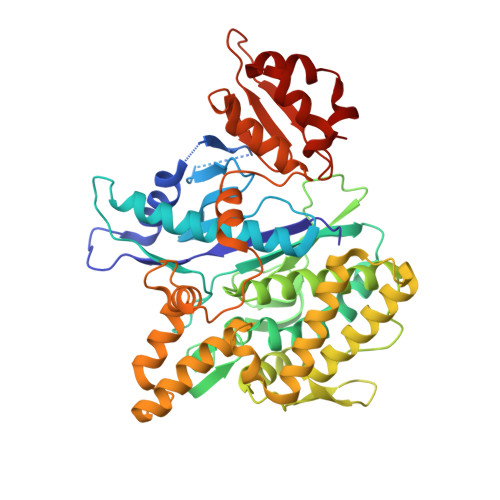
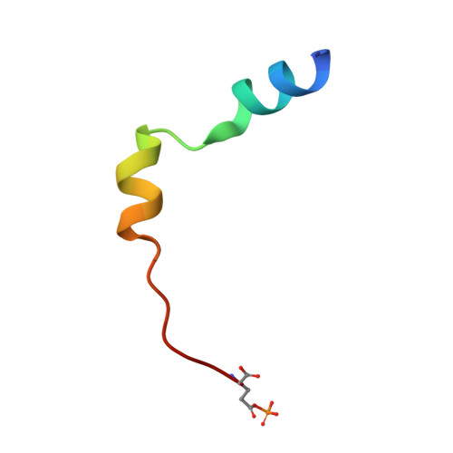
Structures of prokaryotic ubiquitin-like protein Pup in complex with depupylase Dop reveal the mechanism of catalytic phosphate formation.

Cui, H.Muller, A.U.Leibundgut, M.Tian, J.Ban, N.Weber-Ban, E.

(2021) Nat Commun 12: 6635-6635

  • DOI: https://doi.org/10.1038/s41467-021-26848-x
  • Primary Citation Related Structures: 
    7OXV, 7OXY, 7OY3, 7OYF, 7OYH

  • PubMed Abstract: 

    Pupylation is the post-translational modification of lysine side chains with prokaryotic ubiquitin-like protein (Pup) that targets proteins for proteasomal degradation in mycobacteria and other members of Actinobacteria. Pup ligase PafA and depupylase Dop are the two enzymes acting in this pathway. Although they share close structural and sequence homology indicative of a common evolutionary origin, they catalyze opposing reactions. Here, we report a series of high-resolution crystal structures of Dop in different functional states along the reaction pathway, including Pup-bound states in distinct conformations. In combination with biochemical analysis, the structures explain the role of the C-terminal residue of Pup in ATP hydrolysis, the process that generates the catalytic phosphate in the active site, and suggest a role for the Dop-loop as an allosteric sensor for Pup-binding and ATP cleavage.


  • Organizational Affiliation
    • ETH Zurich, Institute of Molecular Biology & Biophysics, CH-8093, Zurich, Switzerland.

Macromolecule Content 

  • Total Structure Weight: 61.95 kDa 
  • Atom Count: 4,595 
  • Modeled Residue Count: 499 
  • Deposited Residue Count: 536 
  • Unique protein chains: 2

Macromolecules

Find similar proteins by:|  3D Structure
Entity ID: 1
MoleculeChains  Sequence LengthOrganismDetailsImage
Depupylase508Acidothermus cellulolyticusMutation(s): 0 
Gene Names: dopAcel_1186
EC: 3.4
UniProt
Find proteins for A0LU48 (Acidothermus cellulolyticus (strain ATCC 43068 / DSM 8971 / 11B))
Explore A0LU48 
Go to UniProtKB:  A0LU48
Entity Groups
Sequence Clusters30% Identity50% Identity70% Identity90% Identity95% Identity100% Identity
UniProt GroupA0LU48
Sequence Annotations
Expand
Reference Sequence
Find similar proteins by:|  3D Structure
Entity ID: 2
MoleculeChains  Sequence LengthOrganismDetailsImage
Prokaryotic ubiquitin-like protein Pup28Acidothermus cellulolyticusMutation(s): 0 
UniProt
Find proteins for A0LU49 (Acidothermus cellulolyticus (strain ATCC 43068 / DSM 8971 / 11B))
Explore A0LU49 
Go to UniProtKB:  A0LU49
Entity Groups
Sequence Clusters30% Identity50% Identity70% Identity90% Identity95% Identity100% Identity
UniProt GroupA0LU49
Sequence Annotations
Expand
Reference Sequence

Small Molecules

Ligands 7 Unique
IDChains Name / Formula / InChI Key2D Diagram3D Interactions
ADP
(Subject of Investigation/LOI)

Query on ADP


Download:Ideal Coordinates CCD File
N [auth A]ADENOSINE-5'-DIPHOSPHATE
C10 H15 N5 O10 P2
XTWYTFMLZFPYCI-KQYNXXCUSA-N
PGE

Query on PGE


Download:Ideal Coordinates CCD File
R [auth A]TRIETHYLENE GLYCOL
C6 H14 O4
ZIBGPFATKBEMQZ-UHFFFAOYSA-N
PEG

Query on PEG


Download:Ideal Coordinates CCD File
Q [auth A]DI(HYDROXYETHYL)ETHER
C4 H10 O3
MTHSVFCYNBDYFN-UHFFFAOYSA-N
EDO

Query on EDO


Download:Ideal Coordinates CCD File
F [auth A]
G [auth A]
H [auth A]
I [auth A]
J [auth A]
F [auth A],
G [auth A],
H [auth A],
I [auth A],
J [auth A],
K [auth A],
L [auth A],
M [auth A]
1,2-ETHANEDIOL
C2 H6 O2
LYCAIKOWRPUZTN-UHFFFAOYSA-N
ACT

Query on ACT


Download:Ideal Coordinates CCD File
O [auth A],
P [auth A]
ACETATE ION
C2 H3 O2
QTBSBXVTEAMEQO-UHFFFAOYSA-M
K
(Subject of Investigation/LOI)

Query on K


Download:Ideal Coordinates CCD File
C [auth A]POTASSIUM ION
K
NPYPAHLBTDXSSS-UHFFFAOYSA-N
MG
(Subject of Investigation/LOI)

Query on MG


Download:Ideal Coordinates CCD File
D [auth A],
E [auth A]
MAGNESIUM ION
Mg
JLVVSXFLKOJNIY-UHFFFAOYSA-N
Modified Residues  1 Unique
IDChains TypeFormula2D DiagramParent
VHF
Query on VHF
B
L-PEPTIDE LINKINGC5 H10 N O7 PGLU

Experimental Data & Validation

Experimental Data

  • Method: X-RAY DIFFRACTION
  • Resolution: 1.78 Å
  • R-Value Free:  0.194 (Depositor), 0.202 (DCC) 
  • R-Value Work:  0.170 (Depositor), 0.176 (DCC) 
  • R-Value Observed: 0.171 (Depositor) 
Space Group: P 31 2 1
Unit Cell:
Length ( Å )Angle ( ˚ )
a = 106.806α = 90
b = 106.806β = 90
c = 108.457γ = 120
Software Package:
Software NamePurpose
XDSdata reduction
Aimlessdata scaling
PHASERphasing
PHENIXrefinement
PDB_EXTRACTdata extraction

Structure Validation

View Full Validation Report


Ligand Structure Quality Assessment 


Entry History 

& Funding Information

Deposition Data

  • Released Date: 2021-12-01 
  • Deposition Author(s): Cui, H.

Funding OrganizationLocationGrant Number
Swiss National Science FoundationSwitzerland31003A_163314

Revision History  (Full details and data files)

  • Version 1.0: 2021-12-01
    Type: Initial release
  • Version 2.0: 2023-03-15
    Changes: Advisory, Atomic model, Author supporting evidence, Data collection, Database references, Derived calculations, Non-polymer description, Polymer sequence, Structure summary
  • Version 2.1: 2024-02-07
    Changes: Data collection, Refinement description