7OTJ | pdb_00007otj

Crystal structure of Pif1 helicase from Candida albicans


Experimental Data Snapshot

  • Method: X-RAY DIFFRACTION
  • Resolution: 2.58 Å
  • R-Value Free: 
    0.242 (Depositor), 0.243 (DCC) 
  • R-Value Work: 
    0.206 (Depositor), 0.207 (DCC) 
  • R-Value Observed: 
    0.208 (Depositor) 

Starting Model: experimental
View more details

wwPDB Validation 3D Report Full Report

Validation slider image for 7OTJ

Ligand Structure Quality Assessment 


This is version 1.1 of the entry. See complete history

Literature

Structural study of the function of Candida Albicans Pif1.

Lu, K.Y.Xin, B.G.Zhang, T.Liu, N.N.Li, D.Rety, S.Xi, X.G.

(2021) Biochem Biophys Res Commun 567: 190-194

  • DOI: https://doi.org/10.1016/j.bbrc.2021.06.050
  • Primary Citation Related Structures: 
    7OTJ

  • PubMed Abstract: 

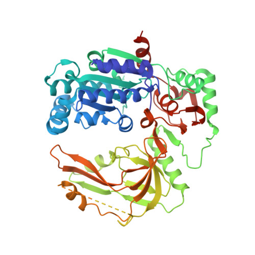
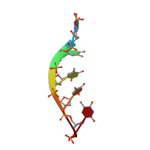
    Pif1 helicases, conserved in eukaryotes, are involved in maintaining genome stability in both the nucleus and mitochondria. Here, we report the crystal structure of a truncated Candida Albicans Pif1 (CaPif1 368-883 ) in complex with ssDNA and an ATP analog. Our results show that the Q-motif is responsible for identifying adenine bases, and CaPif1 preferentially utilizes ATP/dATP during dsDNA unwinding. Although CaPif1 shares structural similarities with Saccharomyces cerevisiae Pif1, CaPif1 can contact the thymidine bases of DNA by hydrogen bonds, whereas ScPif1 cannot. More importantly, the crosslinking and mutant experiments have demonstrated that the conformational change of domain 2B is necessary for CaPif1 to unwind dsDNA. These findings contribute to further the understanding of the unwinding mechanism of Pif1.


  • Organizational Affiliation
    • College of Life Sciences, Northwest A&F University, Yangling, Shaanxi, 712100, China.

Macromolecule Content 

  • Total Structure Weight: 122.33 kDa 
  • Atom Count: 7,347 
  • Modeled Residue Count: 886 
  • Deposited Residue Count: 1,048 
  • Unique protein chains: 1
  • Unique nucleic acid chains: 1

Macromolecules


Find similar proteins by:|  3D Structure
Entity ID: 1
MoleculeChains  Sequence LengthOrganismDetailsImage
ATP-dependent DNA helicase PIF1
A, B
518Candida albicansMutation(s): 0 
EC: 3.6.4.12 (PDB Primary Data), 5.6.2.3 (UniProt)
UniProt
Find proteins for Q59RQ0 (Candida albicans (strain SC5314 / ATCC MYA-2876))
Explore Q59RQ0 
Go to UniProtKB:  Q59RQ0
Entity Groups
Sequence Clusters30% Identity50% Identity70% Identity90% Identity95% Identity100% Identity
UniProt GroupQ59RQ0
Sequence Annotations
Expand
Reference Sequence
Find similar nucleic acids by:  Sequence
Entity ID: 2
MoleculeChains LengthOrganismImage
DNA (5'-D(P*TP*TP*TP*TP*TP*T)-3')
C, D
6Candida albicans
Sequence Annotations
Expand
Reference Sequence

Small Molecules

Ligands 5 Unique
IDChains Name / Formula / InChI Key2D Diagram3D Interactions
ADP
(Subject of Investigation/LOI)

Query on ADP


Download:Ideal Coordinates CCD File
E [auth A],
S [auth B]
ADENOSINE-5'-DIPHOSPHATE
C10 H15 N5 O10 P2
XTWYTFMLZFPYCI-KQYNXXCUSA-N
ALF
(Subject of Investigation/LOI)

Query on ALF


Download:Ideal Coordinates CCD File
F [auth A],
T [auth B]
TETRAFLUOROALUMINATE ION
Al F4
UYOMQIYKOOHAMK-UHFFFAOYSA-J
PO4

Query on PO4


Download:Ideal Coordinates CCD File
O [auth A],
P [auth A],
Q [auth A],
R [auth A],
Y [auth B]
PHOSPHATE ION
O4 P
NBIIXXVUZAFLBC-UHFFFAOYSA-K
K

Query on K


Download:Ideal Coordinates CCD File
H [auth A]
I [auth A]
J [auth A]
K [auth A]
L [auth A]
H [auth A],
I [auth A],
J [auth A],
K [auth A],
L [auth A],
M [auth A],
N [auth A],
V [auth B],
W [auth B],
X [auth B]
POTASSIUM ION
K
NPYPAHLBTDXSSS-UHFFFAOYSA-N
MG
(Subject of Investigation/LOI)

Query on MG


Download:Ideal Coordinates CCD File
G [auth A],
U [auth B]
MAGNESIUM ION
Mg
JLVVSXFLKOJNIY-UHFFFAOYSA-N

Experimental Data & Validation

Experimental Data

  • Method: X-RAY DIFFRACTION
  • Resolution: 2.58 Å
  • R-Value Free:  0.242 (Depositor), 0.243 (DCC) 
  • R-Value Work:  0.206 (Depositor), 0.207 (DCC) 
  • R-Value Observed: 0.208 (Depositor) 
Space Group: C 1 2 1
Unit Cell:
Length ( Å )Angle ( ˚ )
a = 198.973α = 90
b = 79.96β = 117.279
c = 94.11γ = 90
Software Package:
Software NamePurpose
PHENIXrefinement
PHENIXrefinement
XDSdata reduction
Aimlessdata scaling
PHASERphasing

Structure Validation

View Full Validation Report


Ligand Structure Quality Assessment 


Entry History 

Deposition Data

Revision History  (Full details and data files)

  • Version 1.0: 2021-07-07
    Type: Initial release
  • Version 1.1: 2024-01-31
    Changes: Data collection, Database references, Refinement description