Synthesis and biological evaluation of Haspin inhibitors: Kinase inhibitory potency and cellular activity.
Zeinyeh, W., Esvan, Y.J., Josselin, B., Defois, M., Baratte, B., Knapp, S., Chaikuad, A., Anizon, F., Giraud, F., Ruchaud, S., Moreau, P.(2022) Eur J Med Chem 236: 114369-114369
- PubMed: 35447555 Search on PubMed
- DOI: https://doi.org/10.1016/j.ejmech.2022.114369
- Primary Citation Related Structures:
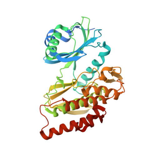
7OPS - PubMed Abstract:
Haspin (haploid germ cell-specific nuclear protein kinase) offers a potential target for the development of new anticancer drugs. Thus, the identification of new inhibitors targeting this protein kinase is of high interest. However, Haspin inhibitors developed to date show a poor selectivity profile over other protein kinases of the human kinome. Here, we identified a new pyridoquinazoline based inhibitor (4), with excellent inhibitory activity and selectivity for Haspin (IC 50 of 50 nM). We describe the structure-activity relationship study including the evaluation of this inhibitor on a large panel of 486 kinases as well as on immortalized or cancer cell lines. In addition, we determined the binding mode of analog 2a in complex with Haspin using X-ray crystallography.
- Université Clermont Auvergne, CNRS, Clermont Auvergne INP, ICCF, F-63000, Clermont-Ferrand, France.
Organizational Affiliation: