7OO4

HaloTag Engineering for Enhanced Fluorogenicity and Kinetics with a Styrylpyridine Dye


Experimental Data Snapshot

  • Method: X-RAY DIFFRACTION
  • Resolution: 2.10 Å
  • R-Value Free: 0.263 
  • R-Value Work: 0.210 
  • R-Value Observed: 0.212 

wwPDB Validation   3D Report Full Report


Ligand Structure Quality Assessment 


This is version 1.3 of the entry. See complete history


Literature

HaloTag Engineering for Enhanced Fluorogenicity and Kinetics with a Styrylpyridium Dye.

Miro-Vinyals, C.Stein, A.Fischer, S.Ward, T.R.Deliz Liang, A.

(2021) Chembiochem 22: 3398-3401

  • DOI: https://doi.org/10.1002/cbic.202100424
  • Primary Citation of Related Structures:  
    7OND, 7OO4

  • PubMed Abstract: 

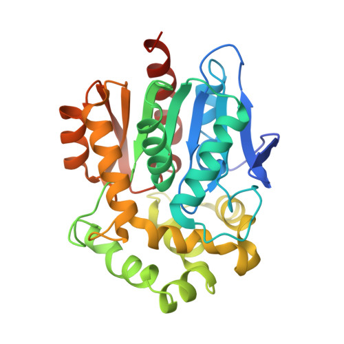
    HaloTag is a small self-labeling protein that is frequently used for creating fluorescent reporters in living cells. The small-molecule dyes used with HaloTag are almost exclusively based on rhodamine scaffolds, which are often expensive and challenging to synthesize. Herein, we report the engineering of HaloTag for use with a chemically accessible, inexpensive fluorophore based on the dimethylamino-styrylpyridium dye. Through directed evolution, the maximum fluorogenicity and the apparent second-order bioconjugation rate constants could be improved up to 4-fold and 42-fold, respectively. One of the top variants, HT-SP5, enabled reliable imaging in mammalian cells, with a 113-fold fluorescence enhancement over the parent protein. Additionally, crystallographic characterization of selected mutants suggests the chemical origin of the fluorescent enhancement. The improved dye system offers a valuable tool for imaging and illustrates the viability of engineering self-labeling proteins for alternative fluorophores.


  • Organizational Affiliation

    Chemistry Department, University of Basel, NCCR Molecular Systems Engineering, 4002, Basel, Switzerland.


Macromolecules
Find similar proteins by:  (by identity cutoff)  |  3D Structure
Entity ID: 1
MoleculeChains Sequence LengthOrganismDetailsImage
Haloalkane dehalogenase300Rhodococcus sp. (in: high G+C Gram-positive bacteria)Mutation(s): 21 
Gene Names: dhaA
EC: 3.8.1.5
UniProt
Find proteins for P0A3G3 (Rhodococcus sp)
Explore P0A3G3 
Go to UniProtKB:  P0A3G3
Entity Groups  
Sequence Clusters30% Identity50% Identity70% Identity90% Identity95% Identity100% Identity
UniProt GroupP0A3G3
Sequence Annotations
Expand
  • Reference Sequence
Small Molecules
Ligands 2 Unique
IDChains Name / Formula / InChI Key2D Diagram3D Interactions
VF2 (Subject of Investigation/LOI)
Query on VF2

Download Ideal Coordinates CCD File 
C [auth A]4-[(E)-2-[1-(7-chloranylheptyl)pyridin-1-ium-4-yl]ethenyl]-N,N-dimethyl-aniline
C22 H30 Cl N2
WTHMXSWCQZGWBE-UHFFFAOYSA-N
CL
Query on CL

Download Ideal Coordinates CCD File 
B [auth A]CHLORIDE ION
Cl
VEXZGXHMUGYJMC-UHFFFAOYSA-M
Experimental Data & Validation

Experimental Data

  • Method: X-RAY DIFFRACTION
  • Resolution: 2.10 Å
  • R-Value Free: 0.263 
  • R-Value Work: 0.210 
  • R-Value Observed: 0.212 
  • Space Group: P 21 21 21
Unit Cell:
Length ( Å )Angle ( ˚ )
a = 52.319α = 90
b = 68.986β = 90
c = 86.02γ = 90
Software Package:
Software NamePurpose
REFMACrefinement
PDB_EXTRACTdata extraction
XDSdata reduction
Aimlessdata scaling
PHASERphasing

Structure Validation

View Full Validation Report


Ligand Structure Quality Assessment 


Entry History 

Deposition Data

Revision History  (Full details and data files)

  • Version 1.0: 2021-07-21
    Type: Initial release
  • Version 1.1: 2021-10-20
    Changes: Data collection, Database references
  • Version 1.2: 2021-12-22
    Changes: Database references
  • Version 1.3: 2024-01-31
    Changes: Data collection, Refinement description