7OO0 | pdb_00007oo0

Mechanosensitive channel MscS solubilized with DDM in open conformation


Experimental Data Snapshot

  • Method: ELECTRON MICROSCOPY
  • Resolution: 3.10 Å
  • Aggregation State: PARTICLE 
  • Reconstruction Method: SINGLE PARTICLE 

Starting Model: experimental
View more details

wwPDB Validation 3D Report Full Report

Validation slider image for 7OO0

This is version 2.0 of the entry. See complete history

Literature

Mechanosensitive channel gating by delipidation.

Flegler, V.J.Rasmussen, A.Borbil, K.Boten, L.Chen, H.A.Deinlein, H.Halang, J.Hellmanzik, K.Loffler, J.Schmidt, V.Makbul, C.Kraft, C.Hedrich, R.Rasmussen, T.Bottcher, B.

(2021) Proc Natl Acad Sci U S A 118

  • DOI: https://doi.org/10.1073/pnas.2107095118
  • Primary Citation Related Structures: 
    7ONJ, 7ONL, 7OO0, 7OO6, 7OO8, 7OOA

  • PubMed Abstract: 

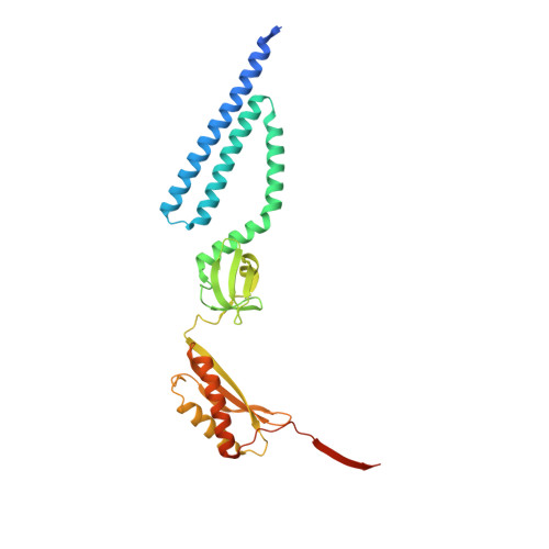
    The mechanosensitive channel of small conductance (MscS) protects bacteria against hypoosmotic shock. It can sense the tension in the surrounding membrane and releases solutes if the pressure in the cell is getting too high. The membrane contacts MscS at sensor paddles, but lipids also leave the membrane and move along grooves between the paddles to reside as far as 15 Å away from the membrane in hydrophobic pockets. One sensing model suggests that a higher tension pulls lipids from the grooves back to the membrane, which triggers gating. However, it is still unclear to what degree this model accounts for sensing and what contribution the direct interaction of the membrane with the channel has. Here, we show that MscS opens when it is sufficiently delipidated by incubation with the detergent dodecyl-β-maltoside or the branched detergent lauryl maltose neopentyl glycol. After addition of detergent-solubilized lipids, it closes again. These results support the model that lipid extrusion causes gating: Lipids are slowly removed from the grooves and pockets by the incubation with detergent, which triggers opening. Addition of lipids in micelles allows lipids to migrate back into the pockets, which closes the channel even in the absence of a membrane. Based on the distribution of the aliphatic chains in the open and closed conformation, we propose that during gating, lipids leave the complex on the cytosolic leaflet at the height of highest lateral tension, while on the periplasmic side, lipids flow into gaps, which open between transmembrane helices.


  • Organizational Affiliation
    • Biocenter and Rudolf-Virchow-Zentrum, Universität Würzburg, 97080 Würzburg, Germany.

Macromolecule Content 

  • Total Structure Weight: 254.19 kDa 
  • Atom Count: 16,016 
  • Modeled Residue Count: 1,834 
  • Deposited Residue Count: 2,058 
  • Unique protein chains: 1

Macromolecules

Find similar proteins by:|  3D Structure
Entity ID: 1
MoleculeChains  Sequence LengthOrganismDetailsImage
Mechanosensitive channel MscS
A, B, C, D, E
A, B, C, D, E, F, G
294Escherichia coliMutation(s): 0 
Membrane Entity: Yes 
UniProt
Find proteins for P0C0S1 (Escherichia coli (strain K12))
Explore P0C0S1 
Go to UniProtKB:  P0C0S1
Entity Groups
Sequence Clusters30% Identity50% Identity70% Identity90% Identity95% Identity100% Identity
UniProt GroupP0C0S1
Sequence Annotations
Expand
Reference Sequence

Small Molecules

Ligands 2 Unique
IDChains Name / Formula / InChI Key2D Diagram3D Interactions
PEE

Query on PEE


Download:Ideal Coordinates CCD File
FB [auth G]
GA [auth D]
H [auth A]
OA [auth E]
Q [auth B]
FB [auth G],
GA [auth D],
H [auth A],
OA [auth E],
Q [auth B],
WA [auth F],
Y [auth C]
1,2-dioleoyl-sn-glycero-3-phosphoethanolamine
C41 H78 N O8 P
MWRBNPKJOOWZPW-NYVOMTAGSA-N
LMT

Query on LMT


Download:Ideal Coordinates CCD File
AA [auth C]
AB [auth F]
BA [auth C]
BB [auth F]
CA [auth C]
AA [auth C],
AB [auth F],
BA [auth C],
BB [auth F],
CA [auth C],
CB [auth G],
DA [auth C],
DB [auth G],
EA [auth C],
EB [auth G],
FA [auth D],
GB [auth G],
HA [auth D],
HB [auth G],
I [auth A],
IA [auth D],
IB [auth G],
J [auth A],
JA [auth D],
JB [auth G],
K [auth A],
KA [auth D],
KB [auth G],
L [auth A],
LA [auth D],
M [auth A],
MA [auth D],
N [auth A],
NA [auth E],
O [auth A],
P [auth B],
PA [auth E],
QA [auth E],
R [auth B],
RA [auth E],
S [auth B],
SA [auth E],
T [auth B],
TA [auth E],
U [auth B],
UA [auth E],
V [auth B],
VA [auth F],
W [auth B],
X [auth C],
XA [auth F],
YA [auth F],
Z [auth C],
ZA [auth F]
DODECYL-BETA-D-MALTOSIDE
C24 H46 O11
NLEBIOOXCVAHBD-QKMCSOCLSA-N

Experimental Data & Validation

Experimental Data

  • Method: ELECTRON MICROSCOPY
  • Resolution: 3.10 Å
  • Aggregation State: PARTICLE 
  • Reconstruction Method: SINGLE PARTICLE 
EM Software:
TaskSoftware PackageVersion
RECONSTRUCTIONRELION3.1
MODEL REFINEMENTPHENIX1.19

Structure Validation

View Full Validation Report


Entry History 

& Funding Information

Deposition Data


Funding OrganizationLocationGrant Number
German Research Foundation (DFG)GermanyBo1150/15-1
German Research Foundation (DFG)GermanyINST 93/903-1 FUGG

Revision History  (Full details and data files)

  • Version 1.0: 2021-09-01
    Type: Initial release
  • Version 2.0: 2024-07-10
    Changes: Atomic model, Data collection, Derived calculations, Non-polymer description, Refinement description, Structure summary