7OM3 | pdb_00007om3

Crystal structure of KOD DNA Polymerase in a binary complex with Hypoxanthine containing template


Experimental Data Snapshot

  • Method: X-RAY DIFFRACTION
  • Resolution: 1.92 Å
  • R-Value Free: 
    0.234 (Depositor), 0.233 (DCC) 
  • R-Value Work: 
    0.183 (Depositor), 0.183 (DCC) 
  • R-Value Observed: 
    0.184 (Depositor) 

Starting Model: experimental
View more details

wwPDB Validation 3D Report Full Report

Validation slider image for 7OM3

This is version 1.3 of the entry. See complete history

Literature

Structural Basis for The Recognition of Deaminated Nucleobases by An Archaeal DNA Polymerase.

Kropp, H.M.Ludmann, S.Diederichs, K.Betz, K.Marx, A.

(2021) Chembiochem 22: 3060-3066

  • DOI: https://doi.org/10.1002/cbic.202100306
  • Primary Citation Related Structures: 
    7OM3, 7OMB, 7OMG

  • PubMed Abstract: 

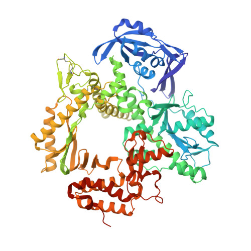
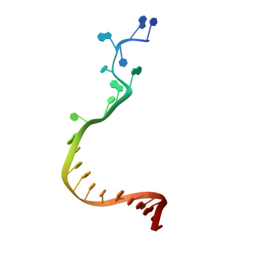
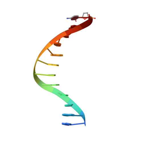
    With increasing temperature, nucleobases in DNA become increasingly damaged by hydrolysis of exocyclic amines. The most prominent damage includes the conversion of cytosine to uracil and adenine to hypoxanthine. These damages are mutagenic and put the integrity of the genome at risk if not repaired appropriately. Several archaea live at elevated temperatures and thus, are exposed to a higher risk of deamination. Earlier studies have shown that DNA polymerases of archaea have the property of sensing deaminated nucleobases in the DNA template and thereby stalling the DNA synthesis during DNA replication providing another layer of DNA damage recognition and repair. However, the structural basis of uracil and hypoxanthine sensing by archaeal B-family DNA polymerases is sparse. Here we report on three new crystal structures of the archaeal B-family DNA polymerase from Thermococcus kodakarensis (KOD) DNA polymerase in complex with primer and template strands that have extended single stranded DNA template 5'-overhangs. These overhangs contain either the canonical nucleobases as well as uracil or hypoxanthine, respectively, and provide unprecedented structural insights into their recognition by archaeal B-family DNA polymerases.


  • Organizational Affiliation
    • Department of Chemistry, University of Konstanz, Universitätsstraße 10, 78457, Konstanz, Germany.

Macromolecule Content 

  • Total Structure Weight: 100.66 kDa 
  • Atom Count: 7,324 
  • Modeled Residue Count: 792 
  • Deposited Residue Count: 807 
  • Unique protein chains: 1
  • Unique nucleic acid chains: 2

Macromolecules


Find similar proteins by:|  3D Structure
Entity ID: 1
MoleculeChains  Sequence LengthOrganismDetailsImage
DNA polymerase,DNA polymerase,DNA polymerase774Thermococcus kodakarensis KOD1Mutation(s): 2 
Gene Names: polTK0001
EC: 2.7.7.7
UniProt
Find proteins for P77933 (Thermococcus kodakarensis (strain ATCC BAA-918 / JCM 12380 / KOD1))
Explore P77933 
Go to UniProtKB:  P77933
Entity Groups
Sequence Clusters30% Identity50% Identity70% Identity90% Identity95% Identity100% Identity
UniProt GroupP77933
Sequence Annotations
Expand
Reference Sequence
Find similar nucleic acids by:  Sequence
Entity ID: 2
MoleculeChains LengthOrganismImage
21nt TemplateB [auth T]21synthetic construct
Sequence Annotations
Expand
Reference Sequence
Find similar nucleic acids by:  Sequence
Entity ID: 3
MoleculeChains LengthOrganismImage
PrimerC [auth P]12synthetic construct
Sequence Annotations
Expand
Reference Sequence

Small Molecules

Ligands 4 Unique
IDChains Name / Formula / InChI Key2D Diagram3D Interactions
PEG

Query on PEG


Download:Ideal Coordinates CCD File
D [auth A]DI(HYDROXYETHYL)ETHER
C4 H10 O3
MTHSVFCYNBDYFN-UHFFFAOYSA-N
GOL

Query on GOL


Download:Ideal Coordinates CCD File
J [auth A]GLYCEROL
C3 H8 O3
PEDCQBHIVMGVHV-UHFFFAOYSA-N
BR

Query on BR


Download:Ideal Coordinates CCD File
H [auth A],
I [auth A]
BROMIDE ION
Br
CPELXLSAUQHCOX-UHFFFAOYSA-M
EDO

Query on EDO


Download:Ideal Coordinates CCD File
E [auth A],
F [auth A],
G [auth A]
1,2-ETHANEDIOL
C2 H6 O2
LYCAIKOWRPUZTN-UHFFFAOYSA-N

Experimental Data & Validation

Experimental Data

  • Method: X-RAY DIFFRACTION
  • Resolution: 1.92 Å
  • R-Value Free:  0.234 (Depositor), 0.233 (DCC) 
  • R-Value Work:  0.183 (Depositor), 0.183 (DCC) 
  • R-Value Observed: 0.184 (Depositor) 
Space Group: P 21 21 2
Unit Cell:
Length ( Å )Angle ( ˚ )
a = 113.356α = 90
b = 142.936β = 90
c = 65.582γ = 90
Software Package:
Software NamePurpose
PHENIXrefinement
PHENIXrefinement
XDSdata reduction
XDSdata scaling
PHENIXphasing

Structure Validation

View Full Validation Report


Entry History 

Deposition Data

Revision History  (Full details and data files)

  • Version 1.0: 2021-10-13
    Type: Initial release
  • Version 1.1: 2021-11-17
    Changes: Data collection, Database references
  • Version 1.2: 2024-01-31
    Changes: Data collection, Refinement description
  • Version 1.3: 2024-11-06
    Changes: Structure summary