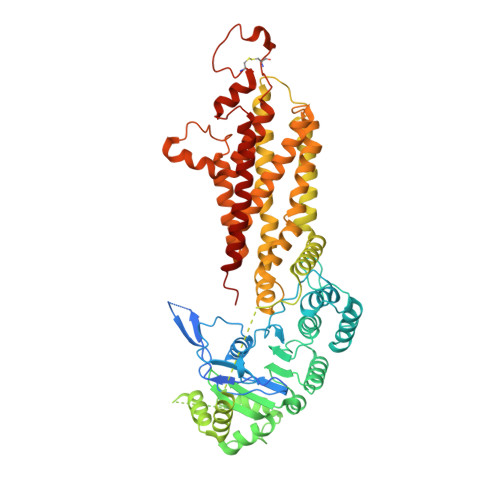
7OJ8

ABCG2 E1S turnover-2 state


Experimental Data Snapshot

  • Method: ELECTRON MICROSCOPY
  • Resolution: 3.40 Å
  • Aggregation State: PARTICLE 
  • Reconstruction Method: SINGLE PARTICLE 

Starting Model: experimental
View more details

wwPDB Validation   3D Report Full Report


This is version 1.2 of the entry. See complete history


Literature

Structures of ABCG2 under turnover conditions reveal a key step in the drug transport mechanism.

Yu, Q.Ni, D.Kowal, J.Manolaridis, I.Jackson, S.M.Stahlberg, H.Locher, K.P.

(2021) Nat Commun 12: 4376-4376

  • DOI: https://doi.org/10.1038/s41467-021-24651-2
  • Primary Citation of Related Structures:  
    7OJ8, 7OJH, 7OJI

  • PubMed Abstract: 

    ABCG2 is a multidrug transporter that affects drug pharmacokinetics and contributes to multidrug resistance of cancer cells. In previously reported structures, the reaction cycle was halted by the absence of substrates or ATP, mutation of catalytic residues, or the presence of small-molecule inhibitors or inhibitory antibodies. Here we present cryo-EM structures of ABCG2 under turnover conditions containing either the endogenous substrate estrone-3-sulfate or the exogenous substrate topotecan. We find two distinct conformational states in which both the transport substrates and ATP are bound. Whereas the state turnover-1 features more widely separated NBDs and an accessible substrate cavity between the TMDs, turnover-2 features semi-closed NBDs and an almost fully occluded substrate cavity. Substrate size appears to control which turnover state is mainly populated. The conformational changes between turnover-1 and turnover-2 states reveal how ATP binding is linked to the closing of the cytoplasmic side of the TMDs. The transition from turnover-1 to turnover-2 is the likely bottleneck or rate-limiting step of the reaction cycle, where the discrimination of substrates and inhibitors occurs.


  • Organizational Affiliation

    Institute of Molecular Biology and Biophysics, Department of Biology, ETH Zürich, Zürich, Switzerland.


Macromolecules
Find similar proteins by:  (by identity cutoff)  |  3D Structure
Entity ID: 1
MoleculeChains Sequence LengthOrganismDetailsImage
Broad substrate specificity ATP-binding cassette transporter ABCG2
A, B
665Homo sapiensMutation(s): 0 
Gene Names: ABCG2ABCPBCRPBCRP1MXR
EC: 7.6.2.2
Membrane Entity: Yes 
UniProt & NIH Common Fund Data Resources
Find proteins for Q9UNQ0 (Homo sapiens)
Explore Q9UNQ0 
Go to UniProtKB:  Q9UNQ0
PHAROS:  Q9UNQ0
GTEx:  ENSG00000118777 
Entity Groups  
Sequence Clusters30% Identity50% Identity70% Identity90% Identity95% Identity100% Identity
UniProt GroupQ9UNQ0
Sequence Annotations
Expand
  • Reference Sequence
Small Molecules
Ligands 3 Unique
IDChains Name / Formula / InChI Key2D Diagram3D Interactions
ATP
Query on ATP

Download Ideal Coordinates CCD File 
C [auth A],
F [auth B]
ADENOSINE-5'-TRIPHOSPHATE
C10 H16 N5 O13 P3
ZKHQWZAMYRWXGA-KQYNXXCUSA-N
CLR
Query on CLR

Download Ideal Coordinates CCD File 
D [auth A],
E [auth A],
H [auth B],
I [auth B]
CHOLESTEROL
C27 H46 O
HVYWMOMLDIMFJA-DPAQBDIFSA-N
FY5 (Subject of Investigation/LOI)
Query on FY5

Download Ideal Coordinates CCD File 
G [auth B]estrone 3-sulfate
C18 H22 O5 S
JKKFKPJIXZFSSB-OVWQWFNUSA-N
Experimental Data & Validation

Experimental Data

  • Method: ELECTRON MICROSCOPY
  • Resolution: 3.40 Å
  • Aggregation State: PARTICLE 
  • Reconstruction Method: SINGLE PARTICLE 
EM Software:
TaskSoftware PackageVersion
MODEL REFINEMENTPHENIX

Structure Validation

View Full Validation Report


Entry History & Funding Information

Deposition Data


Funding OrganizationLocationGrant Number
Swiss National Science FoundationSwitzerlandnot applicable

Revision History  (Full details and data files)

  • Version 1.0: 2021-07-21
    Type: Initial release
  • Version 1.1: 2021-07-28
    Changes: Database references
  • Version 1.2: 2024-10-23
    Changes: Data collection, Database references, Refinement description, Structure summary