7OCJ | pdb_00007ocj

Crystal structure of E.coli LexA in complex with nanobody NbSOS2(Nb14509)


Experimental Data Snapshot

  • Method: X-RAY DIFFRACTION
  • Resolution: 2.70 Å
  • R-Value Free: 
    0.260 (Depositor), 0.252 (DCC) 
  • R-Value Work: 
    0.228 (Depositor), 0.225 (DCC) 

Starting Model: experimental
View more details

wwPDB Validation 3D Report Full Report

Validation slider image for 7OCJ

This is version 1.3 of the entry. See complete history

Literature

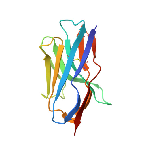
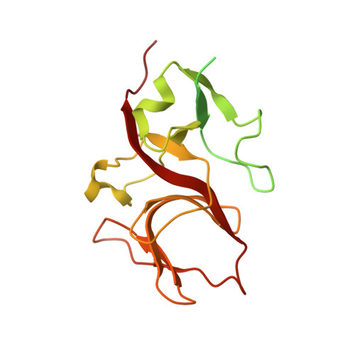
Nanobodies targeting LexA autocleavage disclose a novel suppression strategy of SOS-response pathway.

Maso, L.Vascon, F.Chinellato, M.Goormaghtigh, F.Bellio, P.Campagnaro, E.Van Melderen, L.Ruzzene, M.Pardon, E.Angelini, A.Celenza, G.Steyaert, J.Tondi, D.Cendron, L.

(2022) Structure 30: 1479

  • DOI: https://doi.org/10.1016/j.str.2022.09.004
  • Primary Citation Related Structures: 
    7B5G, 7OCJ, 7ZRA

  • PubMed Abstract: 

    Antimicrobial resistance threatens the eradication of infectious diseases and impairs the efficacy of available therapeutics. The bacterial SOS pathway is a conserved response triggered by genotoxic stresses and represents one of the principal mechanisms that lead to resistance. The RecA recombinase acts as a DNA-damage sensor inducing the autoproteolysis of the transcriptional repressor LexA, thereby derepressing SOS genes that mediate DNA repair, survival to chemotherapy, and hypermutation. The inhibition of such pathway represents a promising strategy for delaying the evolution of antimicrobial resistance. We report the identification, via llama immunization and phage display, of nanobodies that bind LexA with sub-micromolar affinity and block autoproteolysis, repressing SOS response in Escherichia coli. Biophysical characterization of nanobody-LexA complexes revealed that they act by trapping LexA in an inactive conformation and interfering with RecA engagement. Our studies pave the way to the development of new-generation antibiotic adjuvants for the treatment of bacterial infections.


  • Organizational Affiliation
    • Dipartimento di Biologia, Università degli Studi di Padova, Via Ugo Bassi 58/b, 35131 Padova, Italy.

Macromolecule Content 

  • Total Structure Weight: 147.99 kDa 
  • Atom Count: 7,570 
  • Modeled Residue Count: 979 
  • Deposited Residue Count: 1,348 
  • Unique protein chains: 2

Macromolecules

Find similar proteins by:|  3D Structure
Entity ID: 1
MoleculeChains  Sequence LengthOrganismDetailsImage
NbSOS2 (14509)A [auth G],
D [auth E],
E [auth B],
H
115Lama glamaMutation(s): 0 
Find similar proteins by:|  3D Structure
Entity ID: 2
MoleculeChains  Sequence LengthOrganismDetailsImage
LexA repressorB [auth A],
C,
F [auth D],
G [auth F]
222Escherichia coliMutation(s): 0 
Gene Names: lexAEAMG_02325
EC: 3.4.21.88

Small Molecules

Ligands 1 Unique
IDChains Name / Formula / InChI Key2D Diagram3D Interactions
EDO

Query on EDO


Download:Ideal Coordinates CCD File
I [auth A]1,2-ETHANEDIOL
C2 H6 O2
LYCAIKOWRPUZTN-UHFFFAOYSA-N

Experimental Data & Validation

Experimental Data

  • Method: X-RAY DIFFRACTION
  • Resolution: 2.70 Å
  • R-Value Free:  0.260 (Depositor), 0.252 (DCC) 
  • R-Value Work:  0.228 (Depositor), 0.225 (DCC) 
Space Group: P 32 2 1
Unit Cell:
Length ( Å )Angle ( ˚ )
a = 112.672α = 90
b = 112.672β = 90
c = 144.751γ = 120
Software Package:
Software NamePurpose
PHENIXrefinement
PDB_EXTRACTdata extraction
XDSdata reduction
xia2data scaling
MOLREPphasing

Structure Validation

View Full Validation Report


Entry History 

& Funding Information

Deposition Data


Funding OrganizationLocationGrant Number
Italian Ministry of EducationItaly--

Revision History  (Full details and data files)

  • Version 1.0: 2022-10-26
    Type: Initial release
  • Version 1.1: 2022-11-16
    Changes: Database references
  • Version 1.2: 2024-01-31
    Changes: Data collection, Database references, Refinement description
  • Version 1.3: 2024-10-16
    Changes: Structure summary