Unraveling the Mechanics of a Repeat-Protein Nanospring: From Folding of Individual Repeats to Fluctuations of the Superhelix.
Synakewicz, M., Eapen, R.S., Perez-Riba, A., Rowling, P.J.E., Bauer, D., Weissl, A., Fischer, G., Hyvonen, M., Rief, M., Itzhaki, L.S., Stigler, J.(2022) ACS Nano 16: 3895-3905
- PubMed: 35258937 Search on PubMedSearch on PubMed Central
- DOI: https://doi.org/10.1021/acsnano.1c09162
- Primary Citation Related Structures:
7OBI - PubMed Abstract:
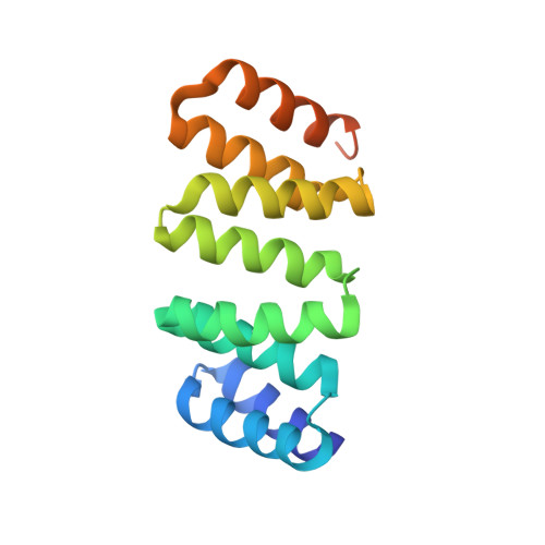
Tandem-repeat proteins comprise small secondary structure motifs that stack to form one-dimensional arrays with distinctive mechanical properties that are proposed to direct their cellular functions. Here, we use single-molecule optical tweezers to study the folding of consensus-designed tetratricopeptide repeats (CTPRs), superhelical arrays of short helix-turn-helix motifs. We find that CTPRs display a spring-like mechanical response in which individual repeats undergo rapid equilibrium fluctuations between partially folded and unfolded conformations. We rationalize the force response using Ising models and dissect the folding pathway of CTPRs under mechanical load, revealing how the repeat arrays form from the center toward both termini simultaneously. Most strikingly, we also directly observe the protein's superhelical tertiary structure in the force signal. Using protein engineering, crystallography, and single-molecule experiments, we show that the superhelical geometry can be altered by carefully placed amino acid substitutions, and we examine how these sequence changes affect intrinsic repeat stability and inter-repeat coupling. Our findings provide the means to dissect and modulate repeat-protein stability and dynamics, which will be essential for researchers to understand the function of natural repeat proteins and to exploit artificial repeats proteins in nanotechnology and biomedical applications.
- Department of Pharmacology, University of Cambridge, Tennis Court Road, Cambridge CB2 1PD, United Kingdom†.
Organizational Affiliation: