7OAJ | pdb_00007oaj

Crystal structure of pseudokinase CASK in complex with compound 7


Experimental Data Snapshot

  • Method: X-RAY DIFFRACTION
  • Resolution: 1.93 Å
  • R-Value Free: 
    0.224 (Depositor), 0.258 (DCC) 
  • R-Value Work: 
    0.195 (Depositor) 
  • R-Value Observed: 
    0.196 (Depositor) 

Starting Model: experimental
View more details

wwPDB Validation 3D Report Full Report

Validation slider image for 7OAJ

Ligand Structure Quality Assessment 


This is version 1.2 of the entry. See complete history

Literature

Design and Development of a Chemical Probe for Pseudokinase Ca 2+ /calmodulin-Dependent Ser/Thr Kinase.

Russ, N.Schroder, M.Berger, B.T.Mandel, S.Aydogan, Y.Mauer, S.Pohl, C.Drewry, D.H.Chaikuad, A.Muller, S.Knapp, S.

(2021) J Med Chem 64: 14358-14376

  • DOI: https://doi.org/10.1021/acs.jmedchem.1c00845
  • Primary Citation Related Structures: 
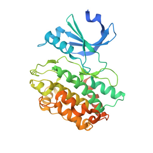
    7OAI, 7OAJ, 7OAK, 7OAL, 7OAM

  • PubMed Abstract: 

    CASK (Ca 2+ /calmodulin-dependent Ser/Thr kinase) is a member of the MAGUK (membrane-associated guanylate kinase) family that functions as neurexin kinases with roles implicated in neuronal synapses and trafficking. The lack of a canonical DFG motif, which is altered to GFG in CASK, led to the classification as a pseudokinase. However, functional studies revealed that CASK can still phosphorylate substrates in the absence of divalent metals. CASK dysfunction has been linked to many diseases, including colorectal cancer, Parkinson's disease, and X-linked mental retardation, suggesting CASK as a potential drug target. Here, we exploited structure-based design for the development of highly potent and selective CASK inhibitors based on 2,4-diaminopyrimidine-5-carboxamides targeting an unusual pocket created by the GFG motif. The presented inhibitor design offers a more general strategy for the development of pseudokinase ligands that harbor unusual sequence motifs. It also provides a first chemical probe for studying the biological roles of CASK.


  • Organizational Affiliation
    • Structural Genomics Consortium (SGC), Buchmann Institute for Life Sciences (BMLS), Goethe University Frankfurt am Main, Max-von-Laue-Str. 15, Frankfurt am Main 60438, Germany.

Macromolecule Content 

  • Total Structure Weight: 161.41 kDa 
  • Atom Count: 10,317 
  • Modeled Residue Count: 1,189 
  • Deposited Residue Count: 1,412 
  • Unique protein chains: 1

Macromolecules

Find similar proteins by:|  3D Structure
Entity ID: 1
MoleculeChains  Sequence LengthOrganismDetailsImage
Peripheral plasma membrane protein CASK
A, B, C, D
353Homo sapiensMutation(s): 0 
Gene Names: CASKLIN2
EC: 2.7.11.1
UniProt & NIH Common Fund Data Resources
Find proteins for O14936 (Homo sapiens)
Explore O14936 
Go to UniProtKB:  O14936
PHAROS:  O14936
GTEx:  ENSG00000147044 
Entity Groups
Sequence Clusters30% Identity50% Identity70% Identity90% Identity95% Identity100% Identity
UniProt GroupO14936
Sequence Annotations
Expand
Reference Sequence

Small Molecules

Ligands 2 Unique
IDChains Name / Formula / InChI Key2D Diagram3D Interactions
V6E
(Subject of Investigation/LOI)

Query on V6E


Download:Ideal Coordinates CCD File
AA [auth D],
E [auth A],
L [auth B],
U [auth C]
4-(cyclopentylamino)-2-[(3,4-dichlorophenyl)methylamino]-N-[3-(2-oxidanylidenepyrrolidin-1-yl)propyl]pyrimidine-5-carboxamide
C24 H30 Cl2 N6 O2
YQQNQEYAEXRCJS-UHFFFAOYSA-N
EDO

Query on EDO


Download:Ideal Coordinates CCD File
BA [auth D]
CA [auth D]
DA [auth D]
F [auth A]
G [auth A]
BA [auth D],
CA [auth D],
DA [auth D],
F [auth A],
G [auth A],
H [auth A],
I [auth A],
J [auth A],
K [auth A],
M [auth B],
N [auth B],
O [auth B],
P [auth B],
Q [auth B],
R [auth B],
S [auth B],
T [auth B],
V [auth C],
W [auth C],
X [auth C],
Y [auth C],
Z [auth C]
1,2-ETHANEDIOL
C2 H6 O2
LYCAIKOWRPUZTN-UHFFFAOYSA-N

Experimental Data & Validation

Experimental Data

  • Method: X-RAY DIFFRACTION
  • Resolution: 1.93 Å
  • R-Value Free:  0.224 (Depositor), 0.258 (DCC) 
  • R-Value Work:  0.195 (Depositor) 
  • R-Value Observed: 0.196 (Depositor) 
Space Group: P 1
Unit Cell:
Length ( Å )Angle ( ˚ )
a = 41.355α = 76.2
b = 66.844β = 89.99
c = 130.203γ = 90
Software Package:
Software NamePurpose
Aimlessdata scaling
REFMACrefinement
PDB_EXTRACTdata extraction
XDSdata reduction
PHASERphasing

Structure Validation

View Full Validation Report


Ligand Structure Quality Assessment 


Entry History 

Deposition Data

Revision History  (Full details and data files)

  • Version 1.0: 2021-05-19
    Type: Initial release
  • Version 1.1: 2021-11-03
    Changes: Data collection, Database references
  • Version 1.2: 2024-01-31
    Changes: Data collection, Refinement description