


Crystal Structure of a Class D Carbapenemase Complexed with Hydrolyzed Oxacillin
Zhou, Q., He, Y., Jin, Y.To be published.
Experimental Data Snapshot
Starting Model: experimental
View more details
Entity ID: 1 | |||||
|---|---|---|---|---|---|
| Molecule | Chains | Sequence Length | Organism | Details | Image |
| Beta-lactamase | A [auth AAA], B [auth BBB], C [auth CCC], D [auth DDD] | 260 | Klebsiella pneumoniae | Mutation(s): 0 Gene Names: bla OXA-48, bla_2, blaOXA-48, G5637_27540, KPE71T_00045, SAMEA3649466_05396 EC: 3.5.2.6 | 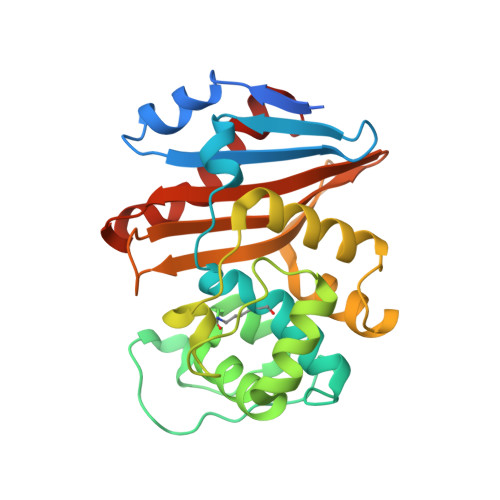 |
UniProt | |||||
Entity Groups | |||||
| Sequence Clusters | 30% Identity50% Identity70% Identity90% Identity95% Identity100% Identity | ||||
| UniProt Group | Q6XEC0 | ||||
Sequence AnnotationsExpand | |||||
Reference Sequence | |||||
| Ligands 5 Unique | |||||
|---|---|---|---|---|---|
| ID | Chains | Name / Formula / InChI Key | 2D Diagram | 3D Interactions | |
| V3H (Subject of Investigation/LOI) Download:Ideal Coordinates CCD File | S [auth CCC], Z [auth DDD] | (2R,4S)-2-[(1R)-2-butoxy-1-[(5-methyl-3-phenyl-1,2-oxazol-4-yl)carbonylamino]-2-oxidanylidene-ethyl]-5,5-dimethyl-1,3-thiazolidin-3-ium-4-carboxylic acid C23 H30 N3 O6 S KTVAJMKGNMTGBA-CMKODMSKSA-O |  | ||
| 0WO (Subject of Investigation/LOI) Download:Ideal Coordinates CCD File | Q [auth CCC], X [auth DDD] | (2R,4S)-2-[(R)-carboxy{[(5-methyl-3-phenyl-1,2-oxazol-4-yl)carbonyl]amino}methyl]-5,5-dimethyl-1,3-thiazolidine-4-carbo
xylic acid C19 H21 N3 O6 S JCHZLBSPYSDECZ-OFQRWUPVSA-N |  | ||
| IOD (Subject of Investigation/LOI) Download:Ideal Coordinates CCD File | AA [auth DDD] BA [auth DDD] F [auth AAA] G [auth AAA] H [auth AAA] | IODIDE ION I XMBWDFGMSWQBCA-UHFFFAOYSA-M |  | ||
| GOL Download:Ideal Coordinates CCD File | E [auth AAA], J [auth BBB], R [auth CCC] | GLYCEROL C3 H8 O3 PEDCQBHIVMGVHV-UHFFFAOYSA-N |  | ||
| 1BO Download:Ideal Coordinates CCD File | K [auth BBB], Y [auth DDD] | 1-BUTANOL C4 H10 O LRHPLDYGYMQRHN-UHFFFAOYSA-N |  | ||
| Modified Residues 1 Unique | |||||
|---|---|---|---|---|---|
| ID | Chains | Type | Formula | 2D Diagram | Parent |
| KCX Query on KCX | A [auth AAA], B [auth BBB], C [auth CCC], D [auth DDD] | L-PEPTIDE LINKING | C7 H14 N2 O4 |  | LYS |
| Length ( Å ) | Angle ( ˚ ) |
|---|---|
| a = 47.053 | α = 90 |
| b = 126.902 | β = 98.289 |
| c = 111.201 | γ = 90 |
| Software Name | Purpose |
|---|---|
| REFMAC | refinement |
| XDS | data reduction |
| XDS | data scaling |
| MOLREP | phasing |
| Funding Organization | Location | Grant Number |
|---|---|---|
| National Science Foundation (NSF, China) | China | 31400663 |


