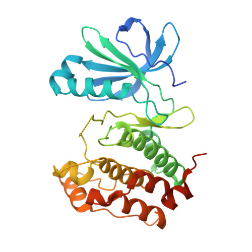
7O2V

AURORA KINASE A IN COMPLEX WITH THE AUR-A/PDK1 INHIBITOR VI8


Experimental Data Snapshot

  • Method: X-RAY DIFFRACTION
  • Resolution: 3.10 Å
  • R-Value Free: 0.329 
  • R-Value Work: 0.272 
  • R-Value Observed: 0.275 

Starting Model: experimental
View more details

wwPDB Validation   3D Report Full Report


Ligand Structure Quality Assessment 


This is version 1.1 of the entry. See complete history


Literature

Development of potent dual PDK1/AurA kinase inhibitors for cancer therapy: Lead-optimization, structural insights, and ADME-Tox profile.

Sestito, S.Bacci, A.Chiarugi, S.Runfola, M.Gado, F.Margheritis, E.Gul, S.Riveiro, M.E.Vazquez, R.Huguet, S.Manera, C.Rezai, K.Garau, G.Rapposelli, S.

(2021) Eur J Med Chem 226: 113895-113895

  • DOI: https://doi.org/10.1016/j.ejmech.2021.113895
  • Primary Citation of Related Structures:  
    7O2V

  • PubMed Abstract: 

    We report the synthesis of novel first-in-class 2-oxindole-based derivatives as dual PDK1-AurA kinase inhibitors as a novel strategy to treat Ewing sarcoma. The most potent compound 12 is suitable for progression to in vivo studies. The specific attributes of 12 included nanomolar inhibitory potency against both phosphoinositide-dependent kinase-1 (PDK1) and Aurora A (AurA) kinase, with acceptable in vitro ADME-Tox properties (cytotoxicity in 2 healthy and 14 hematological and solid cancer cell-lines; inhibition of PDE4C1, SIRT7, HDAC4, HDAC6, HDAC8, HDAC9, AurB, CYP1A2, CYP2C9, CYP2C19, CYP2D6, and hERG). X-ray crystallography and docking studies led to the identification of the key AurA and PDK1/12 interactions. Finally, in vitro drug-intake kinetics and in vivo PK appear to indicate that these compounds are attractive lead-structures for the design and synthesis of PDK1/AurA dual-target molecules to further investigate the in vivo efficacy against Ewing Sarcoma.


  • Organizational Affiliation

    Department of Pharmacy, University of Pisa, 56126, Pisa, Italy.


Macromolecules
Find similar proteins by:  (by identity cutoff)  |  3D Structure
Entity ID: 1
MoleculeChains Sequence LengthOrganismDetailsImage
Aurora kinase A299Homo sapiensMutation(s): 0 
Gene Names: AURKAAIKAIRK1ARK1AURAAYK1BTAKIAK1STK15STK6
EC: 2.7.11.1
UniProt & NIH Common Fund Data Resources
Find proteins for O14965 (Homo sapiens)
Explore O14965 
Go to UniProtKB:  O14965
PHAROS:  O14965
GTEx:  ENSG00000087586 
Entity Groups  
Sequence Clusters30% Identity50% Identity70% Identity90% Identity95% Identity100% Identity
UniProt GroupO14965
Sequence Annotations
Expand
  • Reference Sequence
Small Molecules
Ligands 1 Unique
IDChains Name / Formula / InChI Key2D Diagram3D Interactions
V0K
Query on V0K

Download Ideal Coordinates CCD File 
B [auth A]1-[[3,4-bis(fluoranyl)phenyl]methyl]-~{N}-[(1~{R})-2-[[(3~{E})-3-(1~{H}-imidazol-5-ylmethylidene)-2-oxidanylidene-1~{H}-indol-5-yl]amino]-2-oxidanylidene-1-phenyl-ethyl]-6-methyl-2-oxidanylidene-pyridine-3-carboxamide
C34 H26 F2 N6 O4
KHPDVVDNRUBARQ-WOZRVJTFSA-N
Experimental Data & Validation

Experimental Data

  • Method: X-RAY DIFFRACTION
  • Resolution: 3.10 Å
  • R-Value Free: 0.329 
  • R-Value Work: 0.272 
  • R-Value Observed: 0.275 
  • Space Group: P 61 2 2
Unit Cell:
Length ( Å )Angle ( ˚ )
a = 82.129α = 90
b = 82.129β = 90
c = 166.292γ = 120
Software Package:
Software NamePurpose
MxCuBEdata collection
PHENIXrefinement
MOSFLMdata reduction
Aimlessdata scaling
PHASERphasing

Structure Validation

View Full Validation Report


Ligand Structure Quality Assessment 


Entry History 

Deposition Data

  • Released Date: 2021-10-20 
  • Deposition Author(s): Garau, G.

Revision History  (Full details and data files)

  • Version 1.0: 2021-10-20
    Type: Initial release
  • Version 1.1: 2024-01-31
    Changes: Data collection, Refinement description