7NTC | pdb_00007ntc

Trimeric SARS-CoV-2 spike ectodomain bound to P008_056 Fab


Experimental Data Snapshot

  • Method: ELECTRON MICROSCOPY
  • Resolution: 3.60 Å
  • Aggregation State: PARTICLE 
  • Reconstruction Method: SINGLE PARTICLE 

Starting Model: experimental
View more details

wwPDB Validation 3D Report Full Report

Validation slider image for 7NTC

This is version 1.3 of the entry. See complete history

Literature

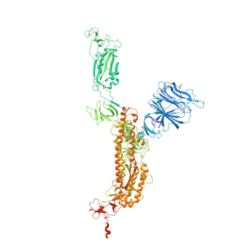
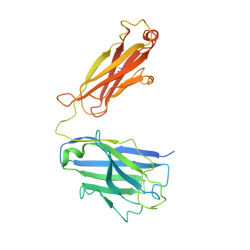
SARS-CoV-2 can recruit a heme metabolite to evade antibody immunity.

Rosa, A.Pye, V.E.Graham, C.Muir, L.Seow, J.Ng, K.W.Cook, N.J.Rees-Spear, C.Parker, E.Dos Santos, M.S.Rosadas, C.Susana, A.Rhys, H.Nans, A.Masino, L.Roustan, C.Christodoulou, E.Ulferts, R.Wrobel, A.G.Short, C.E.Fertleman, M.Sanders, R.W.Heaney, J.Spyer, M.Kjaer, S.Riddell, A.Malim, M.H.Beale, R.MacRae, J.I.Taylor, G.P.Nastouli, E.van Gils, M.J.Rosenthal, P.B.Pizzato, M.McClure, M.O.Tedder, R.S.Kassiotis, G.McCoy, L.E.Doores, K.J.Cherepanov, P.

(2021) Sci Adv 7

  • DOI: https://doi.org/10.1126/sciadv.abg7607
  • Primary Citation Related Structures: 
    7B62, 7NT9, 7NTA, 7NTC

  • PubMed Abstract: 

    The coronaviral spike is the dominant viral antigen and the target of neutralizing antibodies. We show that SARS-CoV-2 spike binds biliverdin and bilirubin, the tetrapyrrole products of heme metabolism, with nanomolar affinity. Using cryo-electron microscopy and x-ray crystallography, we mapped the tetrapyrrole interaction pocket to a deep cleft on the spike N-terminal domain (NTD). At physiological concentrations, biliverdin significantly dampened the reactivity of SARS-CoV-2 spike with immune sera and inhibited a subset of neutralizing antibodies. Access to the tetrapyrrole-sensitive epitope is gated by a flexible loop on the distal face of the NTD. Accompanied by profound conformational changes in the NTD, antibody binding requires relocation of the gating loop, which folds into the cleft vacated by the metabolite. Our results indicate that SARS-CoV-2 spike NTD harbors a dominant epitope, access to which can be controlled by an allosteric mechanism that is regulated through recruitment of a metabolite.


  • Organizational Affiliation
    • Chromatin Structure and Mobile DNA Laboratory, The Francis Crick Institute, London, UK.

Macromolecule Content 

  • Total Structure Weight: 497.49 kDa 
  • Atom Count: 30,005 
  • Modeled Residue Count: 3,698 
  • Deposited Residue Count: 4,346 
  • Unique protein chains: 3

Macromolecules

Find similar proteins by:|  3D Structure
Entity ID: 1
MoleculeChains  Sequence LengthOrganismDetailsImage
Spike glycoprotein
A, B, C
1,287Severe acute respiratory syndrome coronavirus 2Mutation(s): 4 
Gene Names: S2
UniProt
Find proteins for P0DTC2 (Severe acute respiratory syndrome coronavirus 2)
Explore P0DTC2 
Go to UniProtKB:  P0DTC2
Entity Groups
Sequence Clusters30% Identity50% Identity70% Identity90% Identity95% Identity100% Identity
UniProt GroupP0DTC2
Glycosylation
Glycosylation Sites: 18Go to GlyGen: P0DTC2-1
Sequence Annotations
Expand
Reference Sequence
Find similar proteins by:|  3D Structure
Entity ID: 2
MoleculeChains  Sequence LengthOrganismDetailsImage
P008_056 Fab Heavy chainD [auth H]253Homo sapiensMutation(s): 0 
Find similar proteins by:|  3D Structure
Entity ID: 3
MoleculeChains  Sequence LengthOrganismDetailsImage
P008_056 Fab Light chainE [auth L]232Homo sapiensMutation(s): 0 

Oligosaccharides

Help  
Entity ID: 4
MoleculeChains Length2D Diagram GlycosylationD Interactions
2-acetamido-2-deoxy-beta-D-glucopyranose-(1-4)-2-acetamido-2-deoxy-beta-D-glucopyranose
AA [auth a],
BA [auth b],
CA [auth c],
DA [auth d],
F [auth V],
AA [auth a],
BA [auth b],
CA [auth c],
DA [auth d],
F [auth V],
G [auth D],
H [auth E],
I [auth F],
J [auth G],
K [auth I],
L [auth J],
M [auth K],
N [auth M],
O [auth N],
P [auth O],
Q [auth P],
R [auth Q],
S [auth R],
T [auth S],
U [auth T],
V [auth U],
W,
X,
Y,
Z
2N-Glycosylation
Glycosylation Resources
GlyTouCan: G42666HT
GlyCosmos: G42666HT
GlyGen: G42666HT

Small Molecules

Ligands 2 Unique
IDChains Name / Formula / InChI Key2D Diagram3D Interactions
BLA
(Subject of Investigation/LOI)

Query on BLA


Download:Ideal Coordinates CCD File
EA [auth A],
OA [auth B]
BILIVERDINE IX ALPHA
C33 H34 N4 O6
GWZYPXHJIZCRAJ-SRVCBVSDSA-N
NAG

Query on NAG


Download:Ideal Coordinates CCD File
AB [auth C]
BB [auth C]
CB [auth C]
DB [auth C]
EB [auth C]
AB [auth C],
BB [auth C],
CB [auth C],
DB [auth C],
EB [auth C],
FA [auth A],
FB [auth C],
GA [auth A],
GB [auth C],
HA [auth A],
HB [auth C],
IA [auth A],
IB [auth C],
JA [auth A],
KA [auth A],
LA [auth A],
MA [auth A],
NA [auth A],
PA [auth B],
QA [auth B],
RA [auth B],
SA [auth B],
TA [auth B],
UA [auth B],
VA [auth B],
WA [auth B],
XA [auth B],
YA [auth C],
ZA [auth C]
2-acetamido-2-deoxy-beta-D-glucopyranose
C8 H15 N O6
OVRNDRQMDRJTHS-FMDGEEDCSA-N

Experimental Data & Validation

Experimental Data

  • Method: ELECTRON MICROSCOPY
  • Resolution: 3.60 Å
  • Aggregation State: PARTICLE 
  • Reconstruction Method: SINGLE PARTICLE 
EM Software:
TaskSoftware PackageVersion
RECONSTRUCTIONcryoSPARC2
MODEL REFINEMENTPHENIX1.19rc5-4047

Structure Validation

View Full Validation Report


Entry History 

& Funding Information

Deposition Data


Funding OrganizationLocationGrant Number
The Francis Crick InstituteUnited KingdomFC001061

Revision History  (Full details and data files)

  • Version 1.0: 2021-04-28
    Type: Initial release
  • Version 1.1: 2021-05-05
    Changes: Database references
  • Version 1.2: 2021-06-09
    Changes: Database references
  • Version 1.3: 2024-10-23
    Changes: Data collection, Database references, Refinement description, Structure summary