7NRZ | pdb_00007nrz

Crystal structure of malate dehydrogenase from Trypanosoma cruzi


Experimental Data Snapshot

  • Method: X-RAY DIFFRACTION
  • Resolution: 2.60 Å
  • R-Value Free: 
    0.216 (Depositor), 0.217 (DCC) 
  • R-Value Work: 
    0.203 (Depositor), 0.206 (DCC) 
  • R-Value Observed: 
    0.204 (Depositor) 

Starting Model: experimental
View more details

wwPDB Validation 3D Report Full Report

Validation slider image for 7NRZ

This is version 1.1 of the entry. See complete history

Literature

Distinct sequence and structural feature of trypanosoma malate dehydrogenase.

Sonani, R.R.Kurpiewska, K.Lewinski, K.Dubin, G.

(2021) Biochem Biophys Res Commun 557: 288-293

  • DOI: https://doi.org/10.1016/j.bbrc.2021.04.033
  • Primary Citation Related Structures: 
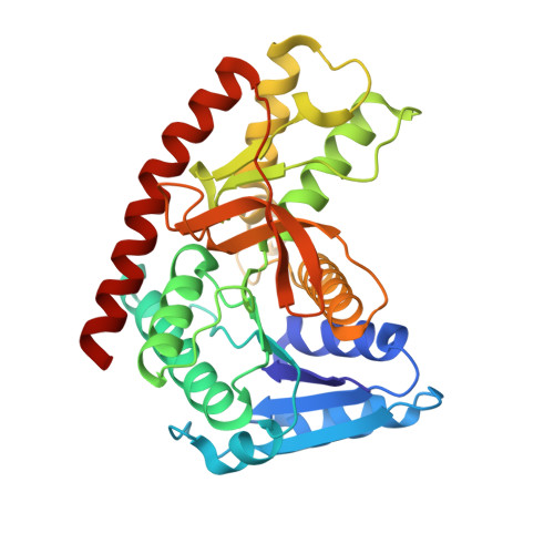
    7NRZ

  • PubMed Abstract: 

    Glycosomal malate dehydrogenase from Trypanosoma cruzi (tcgMDH) catalyzes the oxidation/reduction of malate/oxaloacetate, a crucial step of the glycolytic process occurring in the glycosome of the human parasite. Inhibition of tcgMDH is considered a druggable trait for the development of trypanocidal drugs. Sequence comparison of MDHs from different organisms revealed a distinct insertion of a prolin rich 9-mer (62-KLPPVPRDP-70) in tcgMDH as compared to other eukaryotic MDHs. Crystal structure of tcgMDH is solved here at 2.6 Å resolution with R work /R free values of 0.206/0.216. The tcgMDH forms homo-dimer with the solvation free energy (ΔG o ) gain of -9.77 kcal/mol. The dimeric form is also confirmed in solution by biochemical assays, chemical-crosslinking and dynamic light scattering. The inserted 9-mer adopts a structure of a solvent accessible loop in the vicinity of NAD + binding site. The distinct sequence and structural feature of tcgMDH, revealed in the present report, provides an anchor point for the development of inhibitors specific for tcgMDH, possible trypanocidal agents of the future.


  • Organizational Affiliation
    • Malopolska Centre of Biotechnology, Jagiellonian University, Gronostajowa 7a, 30-387, Krakow, Poland.

Macromolecule Content 

  • Total Structure Weight: 279.01 kDa 
  • Atom Count: 19,867 
  • Modeled Residue Count: 2,584 
  • Deposited Residue Count: 2,584 
  • Unique protein chains: 1

Macromolecules

Find similar proteins by:|  3D Structure
Entity ID: 1
MoleculeChains  Sequence LengthOrganismDetailsImage
Malate dehydrogenase
A, B, C, D, E
A, B, C, D, E, F, G, H
323Trypanosoma cruzi strain CL BrenerMutation(s): 0 
Gene Names: Tc00.1047053506503.69
EC: 1.1.1.37
UniProt
Find proteins for Q4DRD8 (Trypanosoma cruzi (strain CL Brener))
Explore Q4DRD8 
Go to UniProtKB:  Q4DRD8
Entity Groups
Sequence Clusters30% Identity50% Identity70% Identity90% Identity95% Identity100% Identity
UniProt GroupQ4DRD8
Sequence Annotations
Expand
Reference Sequence

Small Molecules

Ligands 5 Unique
IDChains Name / Formula / InChI Key2D Diagram3D Interactions
PG4

Query on PG4


Download:Ideal Coordinates CCD File
BB [auth D],
MB [auth E]
TETRAETHYLENE GLYCOL
C8 H18 O5
UWHCKJMYHZGTIT-UHFFFAOYSA-N
PGE

Query on PGE


Download:Ideal Coordinates CCD File
O [auth A],
Z [auth A]
TRIETHYLENE GLYCOL
C6 H14 O4
ZIBGPFATKBEMQZ-UHFFFAOYSA-N
PEG

Query on PEG


Download:Ideal Coordinates CCD File
AA [auth A],
GA [auth B],
N [auth A],
OA [auth C],
VB [auth F]
DI(HYDROXYETHYL)ETHER
C4 H10 O3
MTHSVFCYNBDYFN-UHFFFAOYSA-N
EDO

Query on EDO


Download:Ideal Coordinates CCD File
AB [auth D]
AC [auth F]
BA [auth B]
BC [auth F]
CA [auth B]
AB [auth D],
AC [auth F],
BA [auth B],
BC [auth F],
CA [auth B],
CB [auth D],
CC [auth F],
DA [auth B],
DB [auth D],
DC [auth F],
EA [auth B],
EB [auth D],
EC [auth G],
FA [auth B],
FB [auth D],
FC [auth G],
GB [auth D],
GC [auth G],
HA [auth B],
HB [auth D],
HC [auth G],
I [auth A],
IA [auth B],
IB [auth D],
IC [auth G],
J [auth A],
JA [auth B],
JB [auth D],
JC [auth G],
K [auth A],
KA [auth B],
KB [auth E],
KC [auth G],
L [auth A],
LA [auth B],
LB [auth E],
LC [auth G],
M [auth A],
MA [auth B],
MC [auth G],
NA [auth B],
NB [auth E],
NC [auth G],
OB [auth E],
P [auth A],
PA [auth C],
PB [auth F],
PC [auth H],
Q [auth A],
QA [auth C],
QB [auth F],
QC [auth H],
R [auth A],
RA [auth C],
RB [auth F],
RC [auth H],
S [auth A],
SA [auth C],
SB [auth F],
SC [auth H],
T [auth A],
TA [auth C],
TB [auth F],
U [auth A],
UA [auth C],
UB [auth F],
V [auth A],
VA [auth C],
W [auth A],
WA [auth C],
WB [auth F],
X [auth A],
XA [auth C],
XB [auth F],
Y [auth A],
YA [auth D],
YB [auth F],
ZA [auth D],
ZB [auth F]
1,2-ETHANEDIOL
C2 H6 O2
LYCAIKOWRPUZTN-UHFFFAOYSA-N
CL

Query on CL


Download:Ideal Coordinates CCD File
OC [auth G]CHLORIDE ION
Cl
VEXZGXHMUGYJMC-UHFFFAOYSA-M

Experimental Data & Validation

Experimental Data

  • Method: X-RAY DIFFRACTION
  • Resolution: 2.60 Å
  • R-Value Free:  0.216 (Depositor), 0.217 (DCC) 
  • R-Value Work:  0.203 (Depositor), 0.206 (DCC) 
  • R-Value Observed: 0.204 (Depositor) 
Space Group: P 21 21 21
Unit Cell:
Length ( Å )Angle ( ˚ )
a = 80.04α = 90
b = 148.331β = 90
c = 251.8γ = 90
Software Package:
Software NamePurpose
PHENIXrefinement
PDB_EXTRACTdata extraction
XDSdata reduction
Aimlessdata scaling
PHASERphasing

Structure Validation

View Full Validation Report


Entry History 

& Funding Information

Deposition Data


Funding OrganizationLocationGrant Number
Polish National Science CentrePoland2017/26/M/NZ1/00797
Foundation for Polish SciencePolandTEAM TECH CORE FACILITY/2017-4/6

Revision History  (Full details and data files)

  • Version 1.0: 2022-02-16
    Type: Initial release
  • Version 1.1: 2024-01-31
    Changes: Data collection, Refinement description