7NRF

Crystal structure of E.coli BamA beta-barrel in complex with darobactin (crystal form 2)


Experimental Data Snapshot

  • Method: X-RAY DIFFRACTION
  • Resolution: 2.20 Å
  • R-Value Free: 0.232 
  • R-Value Work: 0.218 
  • R-Value Observed: 0.219 

wwPDB Validation   3D Report Full Report


Ligand Structure Quality Assessment 


This is version 1.3 of the entry. See complete history


Literature

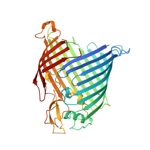
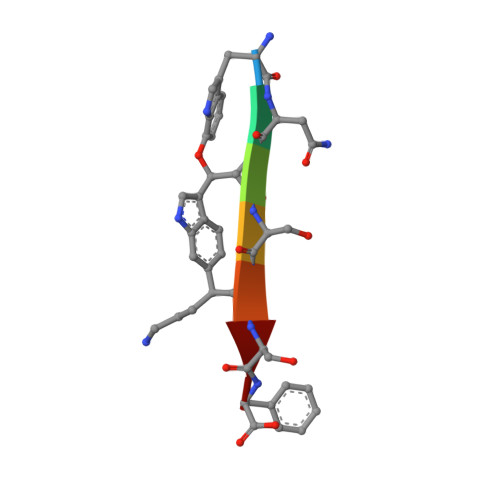
The antibiotic darobactin mimics a beta-strand to inhibit outer membrane insertase.

Kaur, H.Jakob, R.P.Marzinek, J.K.Green, R.Imai, Y.Bolla, J.R.Agustoni, E.Robinson, C.V.Bond, P.J.Lewis, K.Maier, T.Hiller, S.

(2021) Nature 593: 125-129

  • DOI: https://doi.org/10.1038/s41586-021-03455-w
  • Primary Citation of Related Structures:  
    7NRE, 7NRF, 7NRI

  • PubMed Abstract: 

    Antibiotics that target Gram-negative bacteria in new ways are needed to resolve the antimicrobial resistance crisis 1-3 . Gram-negative bacteria are protected by an additional outer membrane, rendering proteins on the cell surface attractive drug targets 4,5 . The natural compound darobactin targets the bacterial insertase BamA 6 -the central unit of the essential BAM complex, which facilitates the folding and insertion of outer membrane proteins 7-13 . BamA lacks a typical catalytic centre, and it is not obvious how a small molecule such as darobactin might inhibit its function. Here we resolve the mode of action of darobactin at the atomic level using a combination of cryo-electron microscopy, X-ray crystallography, native mass spectrometry, in vivo experiments and molecular dynamics simulations. Two cyclizations pre-organize the darobactin peptide in a rigid β-strand conformation. This creates a mimic of the recognition signal of native substrates with a superior ability to bind to the lateral gate of BamA. Upon binding, darobactin replaces a lipid molecule from the lateral gate to use the membrane environment as an extended binding pocket. Because the interaction between darobactin and BamA is largely mediated by backbone contacts, it is particularly robust against potential resistance mutations. Our results identify the lateral gate as a functional hotspot in BamA and will allow the rational design of antibiotics that target this bacterial Achilles heel.


  • Organizational Affiliation

    Biozentrum, University of Basel, Basel, Switzerland.


Macromolecules
Find similar proteins by:  (by identity cutoff)  |  3D Structure
Entity ID: 1
MoleculeChains Sequence LengthOrganismDetailsImage
Outer membrane protein assembly factor BamA411Escherichia coli O157:H7Mutation(s): 0 
Gene Names: bamAyaeTZ0188ECs0179
Membrane Entity: Yes 
UniProt
Find proteins for P0A940 (Escherichia coli (strain K12))
Explore P0A940 
Go to UniProtKB:  P0A940
Entity Groups  
Sequence Clusters30% Identity50% Identity70% Identity90% Identity95% Identity100% Identity
UniProt GroupP0A940
Sequence Annotations
Expand
  • Reference Sequence

Find similar proteins by:  Sequence   |   3D Structure  

Entity ID: 2
MoleculeChains Sequence LengthOrganismDetailsImage
Darobactin7synthetic constructMutation(s): 0 
Sequence Annotations
Expand
  • Reference Sequence
Small Molecules
Ligands 2 Unique
IDChains Name / Formula / InChI Key2D Diagram3D Interactions
C8E
Query on C8E

Download Ideal Coordinates CCD File 
D [auth A]
E [auth A]
F [auth A]
G [auth A]
H [auth A]
D [auth A],
E [auth A],
F [auth A],
G [auth A],
H [auth A],
I [auth A],
J [auth A],
K [auth A]
(HYDROXYETHYLOXY)TRI(ETHYLOXY)OCTANE
C16 H34 O5
FEOZZFHAVXYAMB-UHFFFAOYSA-N
MG
Query on MG

Download Ideal Coordinates CCD File 
C [auth A]MAGNESIUM ION
Mg
JLVVSXFLKOJNIY-UHFFFAOYSA-N
Modified Residues  1 Unique
IDChains TypeFormula2D DiagramParent
UX8
Query on UX8
B
L-PEPTIDE LINKINGC11 H12 N2 O3TRP
Biologically Interesting Molecules (External Reference) 1 Unique
Experimental Data & Validation

Experimental Data

  • Method: X-RAY DIFFRACTION
  • Resolution: 2.20 Å
  • R-Value Free: 0.232 
  • R-Value Work: 0.218 
  • R-Value Observed: 0.219 
  • Space Group: I 1 2 1
Unit Cell:
Length ( Å )Angle ( ˚ )
a = 79.61α = 90
b = 79.814β = 106.42
c = 89.346γ = 90
Software Package:
Software NamePurpose
PHENIXrefinement
PDB_EXTRACTdata extraction
XDSdata reduction
Aimlessdata scaling
PHASERphasing

Structure Validation

View Full Validation Report


Ligand Structure Quality Assessment 


Entry History 

Deposition Data

Revision History  (Full details and data files)

  • Version 1.0: 2021-04-21
    Type: Initial release
  • Version 1.1: 2021-04-28
    Changes: Database references
  • Version 1.2: 2021-05-19
    Changes: Database references
  • Version 1.3: 2024-01-31
    Changes: Data collection, Database references, Derived calculations, Refinement description