


Galectin-1 in complex with 4-Amino-6-chloro-1,3-benzenedisulfonamide
Grimm, C., Bechold, J., Seibel, J.To be published.
Experimental Data Snapshot
Starting Model: experimental
View more details
Entity ID: 1 | |||||
|---|---|---|---|---|---|
| Molecule | Chains | Sequence Length | Organism | Details | Image |
| Galectin-1 | 133 | Homo sapiens | Mutation(s): 0 Gene Names: LGALS1 | 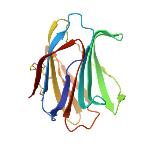 | |
UniProt & NIH Common Fund Data Resources | |||||
PHAROS: P09382 GTEx: ENSG00000100097 | |||||
Entity Groups | |||||
| Sequence Clusters | 30% Identity50% Identity70% Identity90% Identity95% Identity100% Identity | ||||
| UniProt Group | P09382 | ||||
Sequence AnnotationsExpand | |||||
Reference Sequence | |||||
| Ligands 2 Unique | |||||
|---|---|---|---|---|---|
| ID | Chains | Name / Formula / InChI Key | 2D Diagram | 3D Interactions | |
| I7B (Subject of Investigation/LOI) Download:Ideal Coordinates CCD File | D [auth B] | 4-AMINO-6-CHLOROBENZENE-1,3-DISULFONAMIDE C6 H8 Cl N3 O4 S2 IHJCXVZDYSXXFT-UHFFFAOYSA-N |  | ||
| DMS Download:Ideal Coordinates CCD File | C [auth A] E [auth B] F [auth B] G [auth B] H [auth B] | DIMETHYL SULFOXIDE C2 H6 O S IAZDPXIOMUYVGZ-UHFFFAOYSA-N |  | ||
| Modified Residues 1 Unique | |||||
|---|---|---|---|---|---|
| ID | Chains | Type | Formula | 2D Diagram | Parent |
| CME Query on CME | A, B | L-PEPTIDE LINKING | C5 H11 N O3 S2 |  | CYS |
| Length ( Å ) | Angle ( ˚ ) |
|---|---|
| a = 43.319 | α = 90 |
| b = 58.475 | β = 90 |
| c = 112.04 | γ = 90 |
| Software Name | Purpose |
|---|---|
| PHENIX | refinement |
| PHENIX | refinement |
| XDS | data reduction |
| XDS | data scaling |
| PHASER | phasing |


