7NLD | pdb_00007nld

Structure of human Programmed cell death 1 ligand 1 (PD-L1) with low molecular mass inhibitor


Experimental Data Snapshot

  • Method: X-RAY DIFFRACTION
  • Resolution: 2.30 Å
  • R-Value Free: 
    0.294 (Depositor), 0.297 (DCC) 
  • R-Value Work: 
    0.243 (Depositor), 0.245 (DCC) 
  • R-Value Observed: 
    0.244 (Depositor) 

Starting Model: experimental
View more details

wwPDB Validation 3D Report Full Report

Validation slider image for 7NLD

Ligand Structure Quality Assessment 


This is version 1.3 of the entry. See complete history

Literature

Terphenyl-Based Small-Molecule Inhibitors of Programmed Cell Death-1/Programmed Death-Ligand 1 Protein-Protein Interaction.

Muszak, D.Surmiak, E.Plewka, J.Magiera-Mularz, K.Kocik-Krol, J.Musielak, B.Sala, D.Kitel, R.Stec, M.Weglarczyk, K.Siedlar, M.Domling, A.Skalniak, L.Holak, T.A.

(2021) J Med Chem 64: 11614-11636

  • DOI: https://doi.org/10.1021/acs.jmedchem.1c00957
  • Primary Citation Related Structures: 
    6R3K, 7NLD

  • PubMed Abstract: 

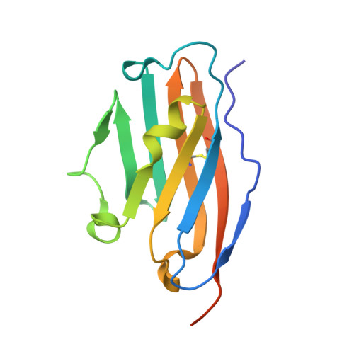
    We describe a new class of potent PD-L1/PD-1 inhibitors based on a terphenyl scaffold that is derived from the rigidified biphenyl-inspired structure. Using in silico docking, we designed and then experimentally demonstrated the effectiveness of the terphenyl-based scaffolds in inhibiting PD-1/PD-L1 complex formation using various biophysical and biochemical techniques. We also present a high-resolution structure of the complex of PD-L1 with one of our most potent inhibitors to identify key PD-L1/inhibitor interactions at the molecular level. In addition, we show the efficacy of our most potent inhibitors in activating the antitumor response using primary human immune cells from healthy donors.


  • Organizational Affiliation
    • Department of Organic Chemistry, Faculty of Chemistry, Jagiellonian University, Gronostajowa 2, 30-387 Krakow, Poland.

Macromolecule Content 

  • Total Structure Weight: 89.58 kDa 
  • Atom Count: 5,354 
  • Modeled Residue Count: 696 
  • Deposited Residue Count: 768 
  • Unique protein chains: 1

Macromolecules

Find similar proteins by:|  3D Structure
Entity ID: 1
MoleculeChains  Sequence LengthOrganismDetailsImage
Programmed cell death 1 ligand 1128Homo sapiensMutation(s): 0 
Gene Names: CD274B7H1PDCD1L1PDCD1LG1PDL1
UniProt & NIH Common Fund Data Resources
Find proteins for Q9NZQ7 (Homo sapiens)
Explore Q9NZQ7 
Go to UniProtKB:  Q9NZQ7
PHAROS:  Q9NZQ7
GTEx:  ENSG00000120217 
Entity Groups
Sequence Clusters30% Identity50% Identity70% Identity90% Identity95% Identity100% Identity
UniProt GroupQ9NZQ7
Sequence Annotations
Expand
Reference Sequence

Experimental Data & Validation

Experimental Data

  • Method: X-RAY DIFFRACTION
  • Resolution: 2.30 Å
  • R-Value Free:  0.294 (Depositor), 0.297 (DCC) 
  • R-Value Work:  0.243 (Depositor), 0.245 (DCC) 
  • R-Value Observed: 0.244 (Depositor) 
Space Group: P 1 21 1
Unit Cell:
Length ( Å )Angle ( ˚ )
a = 67.087α = 90
b = 75.255β = 96.29
c = 74.354γ = 90
Software Package:
Software NamePurpose
PHENIXrefinement
PDB_EXTRACTdata extraction
XDSdata reduction
SCALAdata scaling
PHASERphasing

Structure Validation

View Full Validation Report


Ligand Structure Quality Assessment 


Entry History 

Deposition Data

Revision History  (Full details and data files)

  • Version 1.0: 2021-08-11
    Type: Initial release
  • Version 1.1: 2021-08-25
    Changes: Database references
  • Version 1.2: 2024-01-31
    Changes: Data collection, Refinement description
  • Version 1.3: 2024-11-13
    Changes: Structure summary