7NF8 | pdb_00007nf8

Ovine (b0,+AT-rBAT)2 hetero-tetramer, asymmetric unit, rigid-body fitted


Experimental Data Snapshot

  • Method: ELECTRON MICROSCOPY
  • Resolution: 2.83 Å
  • Aggregation State: PARTICLE 
  • Reconstruction Method: SINGLE PARTICLE 

wwPDB Validation 3D Report Full Report

Validation slider image for 7NF8

This is version 1.4 of the entry. See complete history

Literature

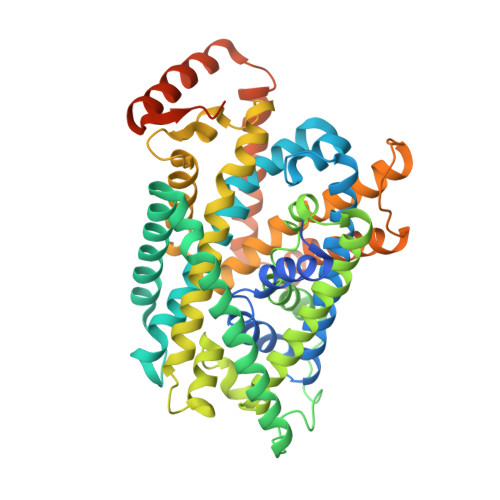
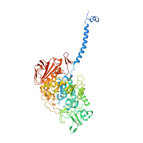
Ca 2+ -mediated higher-order assembly of heterodimers in amino acid transport system b 0,+ biogenesis and cystinuria.

Lee, Y.Wiriyasermkul, P.Kongpracha, P.Moriyama, S.Mills, D.J.Kuhlbrandt, W.Nagamori, S.

(2022) Nat Commun 13: 2708-2708

  • DOI: https://doi.org/10.1038/s41467-022-30293-9
  • Primary Citation Related Structures: 
    7NF6, 7NF7, 7NF8

  • PubMed Abstract: 

    Cystinuria is a genetic disorder characterized by overexcretion of dibasic amino acids and cystine, causing recurrent kidney stones and kidney failure. Mutations of the regulatory glycoprotein rBAT and the amino acid transporter b 0,+ AT, which constitute system b 0,+ , are linked to type I and non-type I cystinuria respectively and they exhibit distinct phenotypes due to protein trafficking defects or catalytic inactivation. Here, using electron cryo-microscopy and biochemistry, we discover that Ca 2+ mediates higher-order assembly of system b 0,+ . Ca 2+ stabilizes the interface between two rBAT molecules, leading to super-dimerization of b 0,+ AT-rBAT, which in turn facilitates N-glycan maturation and protein trafficking. A cystinuria mutant T216M and mutations of the Ca 2+ site of rBAT cause the loss of higher-order assemblies, resulting in protein trapping at the ER and the loss of function. These results provide the molecular basis of system b 0,+ biogenesis and type I cystinuria and serve as a guide to develop new therapeutic strategies against it. More broadly, our findings reveal an unprecedented link between transporter oligomeric assembly and protein-trafficking diseases.


  • Organizational Affiliation
    • Department of Structural Biology, Max Planck Institute of Biophysics, 60438, Frankfurt, Germany. yongchan.lee@biophys.mpg.de.

Macromolecule Content 

  • Total Structure Weight: 138.38 kDa 
  • Atom Count: 8,817 
  • Modeled Residue Count: 1,078 
  • Deposited Residue Count: 1,201 
  • Unique protein chains: 2

Macromolecules

Find similar proteins by:|  3D Structure
Entity ID: 1
MoleculeChains  Sequence LengthOrganismDetailsImage
B(0,+)-type amino acid transporter 1A [auth B]515Ovis ariesMutation(s): 0 
Gene Names: SLC7A9
Find similar proteins by:|  3D Structure
Entity ID: 2
MoleculeChains  Sequence LengthOrganismDetailsImage
neutral and basic amino acid transport protein rBATB [auth A]686Ovis ariesMutation(s): 0 
Gene Names: SLC3A1
Membrane Entity: Yes 
UniProt
Find proteins for A0A6P7DVK7 (Ovis aries)
Explore A0A6P7DVK7 
Go to UniProtKB:  A0A6P7DVK7
Entity Groups
Sequence Clusters30% Identity50% Identity70% Identity90% Identity95% Identity100% Identity
UniProt GroupA0A6P7DVK7
Glycosylation
Glycosylation Sites: 6
Sequence Annotations
Expand
Reference Sequence

Oligosaccharides

Help  
Entity ID: 3
MoleculeChains Length2D Diagram GlycosylationD Interactions
2-acetamido-2-deoxy-beta-D-glucopyranose-(1-4)-2-acetamido-2-deoxy-beta-D-glucopyranose
C, D
2N-Glycosylation
Glycosylation Resources
GlyTouCan: G42666HT
GlyCosmos: G42666HT
GlyGen: G42666HT

Small Molecules

Ligands 4 Unique
IDChains Name / Formula / InChI Key2D Diagram3D Interactions
LBN

Query on LBN


Download:Ideal Coordinates CCD File
G [auth A]1-palmitoyl-2-oleoyl-sn-glycero-3-phosphocholine
C42 H82 N O8 P
WTJKGGKOPKCXLL-VYOBOKEXSA-N
CLR

Query on CLR


Download:Ideal Coordinates CCD File
E [auth B],
F [auth B]
CHOLESTEROL
C27 H46 O
HVYWMOMLDIMFJA-DPAQBDIFSA-N
NAG

Query on NAG


Download:Ideal Coordinates CCD File
I [auth A],
J [auth A],
K [auth A],
L [auth A]
2-acetamido-2-deoxy-beta-D-glucopyranose
C8 H15 N O6
OVRNDRQMDRJTHS-FMDGEEDCSA-N
CA
(Subject of Investigation/LOI)

Query on CA


Download:Ideal Coordinates CCD File
H [auth A]CALCIUM ION
Ca
BHPQYMZQTOCNFJ-UHFFFAOYSA-N

Experimental Data & Validation

Experimental Data

  • Method: ELECTRON MICROSCOPY
  • Resolution: 2.83 Å
  • Aggregation State: PARTICLE 
  • Reconstruction Method: SINGLE PARTICLE 
EM Software:
TaskSoftware PackageVersion
MODEL REFINEMENTPHENIX

Structure Validation

View Full Validation Report


Entry History 

& Funding Information

Deposition Data


Funding OrganizationLocationGrant Number
Max Planck SocietyGermany--
Human Frontier Science Program (HFSP)--

Revision History  (Full details and data files)

  • Version 1.0: 2022-01-19
    Type: Initial release
  • Version 1.1: 2022-06-01
    Changes: Database references, Derived calculations
  • Version 1.2: 2022-06-08
    Changes: Database references
  • Version 1.3: 2024-10-16
    Changes: Data collection, Database references, Structure summary
  • Version 1.4: 2025-07-09
    Changes: Data collection