7NCV | pdb_00007ncv

Crystal structure of reduced glutaredoxin 2 from Chlamydomonas reinhardtii


Experimental Data Snapshot

  • Method: X-RAY DIFFRACTION
  • Resolution: 1.50 Å
  • R-Value Free: 
    0.180 (Depositor), 0.182 (DCC) 
  • R-Value Work: 
    0.153 (Depositor), 0.155 (DCC) 
  • R-Value Observed: 
    0.154 (Depositor) 

Starting Model: experimental
View more details

wwPDB Validation 3D Report Full Report

Validation slider image for 7NCV

This is version 1.3 of the entry. See complete history

Literature

Atypical Iron-Sulfur Cluster Binding, Redox Activity and Structural Properties of Chlamydomonas reinhardtii Glutaredoxin 2.

Roret, T.Zhang, B.Moseler, A.Dhalleine, T.Gao, X.H.Couturier, J.Lemaire, S.D.Didierjean, C.Johnson, M.K.Rouhier, N.

(2021) Antioxidants (Basel) 10

  • DOI: https://doi.org/10.3390/antiox10050803
  • Primary Citation Related Structures: 
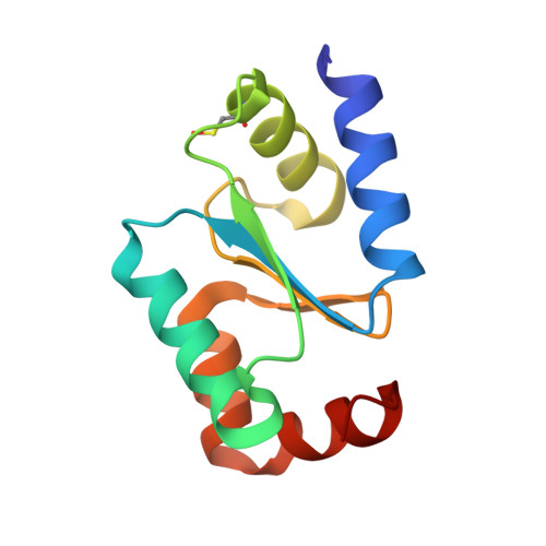
    7NCV, 7NCW

  • PubMed Abstract: 

    Glutaredoxins (GRXs) are thioredoxin superfamily members exhibiting thiol-disulfide oxidoreductase activity and/or iron-sulfur (Fe-S) cluster binding capacities. These properties are determined by specific structural factors. In this study, we examined the capacity of the class I Chlamydomonas reinhardtii GRX2 recombinant protein to catalyze both protein glutathionylation and deglutathionylation reactions using a redox sensitive fluorescent protein as a model protein substrate. We observed that the catalytic cysteine of the CPYC active site motif of GRX2 was sufficient for catalyzing both reactions in the presence of glutathione. Unexpectedly, spectroscopic characterization of the protein purified under anaerobiosis showed the presence of a [2Fe-2S] cluster despite having a presumably inadequate active site signature, based on past mutational analyses. The spectroscopic characterization of cysteine mutated variants together with modeling of the Fe-S cluster-bound GRX homodimer from the structure of an apo-GRX2 indicate the existence of an atypical Fe-S cluster environment and ligation mode. Overall, the results further delineate the biochemical and structural properties of conventional GRXs, pointing to the existence of multiple factors more complex than anticipated, sustaining the capacity of these proteins to bind Fe-S clusters.


  • Organizational Affiliation
    • Université de Lorraine, INRAE, IAM, F-54000 Nancy, France.

Macromolecule Content 

  • Total Structure Weight: 11.46 kDa 
  • Atom Count: 973 
  • Modeled Residue Count: 107 
  • Deposited Residue Count: 107 
  • Unique protein chains: 1

Macromolecules

Find similar proteins by:|  3D Structure
Entity ID: 1
MoleculeChains  Sequence LengthOrganismDetailsImage
Glutaredoxin, CPYC type107Chlamydomonas reinhardtiiMutation(s): 0 
Gene Names: GRX2CHLRE_12g550400v5CHLREDRAFT_195611
UniProt
Find proteins for A8IYH1 (Chlamydomonas reinhardtii)
Explore A8IYH1 
Go to UniProtKB:  A8IYH1
Entity Groups
Sequence Clusters30% Identity50% Identity70% Identity90% Identity95% Identity100% Identity
UniProt GroupA8IYH1
Sequence Annotations
Expand
Reference Sequence

Experimental Data & Validation

Experimental Data

  • Method: X-RAY DIFFRACTION
  • Resolution: 1.50 Å
  • R-Value Free:  0.180 (Depositor), 0.182 (DCC) 
  • R-Value Work:  0.153 (Depositor), 0.155 (DCC) 
  • R-Value Observed: 0.154 (Depositor) 
Space Group: P 32 2 1
Unit Cell:
Length ( Å )Angle ( ˚ )
a = 45.648α = 90
b = 45.648β = 90
c = 104.667γ = 120
Software Package:
Software NamePurpose
XDSdata reduction
SCALAdata scaling
Cootmodel building
MOLREPphasing
PHENIXrefinement

Structure Validation

View Full Validation Report


Entry History 

Deposition Data

Revision History  (Full details and data files)

  • Version 1.0: 2021-05-26
    Type: Initial release
  • Version 1.1: 2021-06-16
    Changes: Database references
  • Version 1.2: 2024-01-31
    Changes: Data collection, Database references, Refinement description
  • Version 1.3: 2024-10-23
    Changes: Structure summary