7NBI | pdb_00007nbi

Crystal structure of a monomeric FLT3 Ligand variant


Experimental Data Snapshot

  • Method: X-RAY DIFFRACTION
  • Resolution: 1.65 Å
  • R-Value Free: 
    0.203 (Depositor), 0.203 (DCC) 
  • R-Value Work: 
    0.164 (Depositor), 0.165 (DCC) 
  • R-Value Observed: 
    0.166 (Depositor) 

Starting Model: experimental
View more details

wwPDB Validation 3D Report Full Report

Validation slider image for 7NBI

This is version 1.2 of the entry. See complete history

Literature

Engineering and crystal structure of a monomeric FLT3 ligand variant.

Pannecoucke, E.Raes, L.Savvides, S.N.

(2021) Acta Crystallogr F Struct Biol Commun 77: 121-127

  • DOI: https://doi.org/10.1107/S2053230X21003289
  • Primary Citation Related Structures: 
    7NBI

  • PubMed Abstract: 

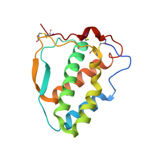
    The overarching paradigm for the activation of class III and V receptor tyrosine kinases (RTKs) prescribes cytokine-mediated dimerization of the receptor ectodomains and homotypic receptor-receptor interactions. However, structural studies have shown that the hematopoietic receptor FLT3, a class III RTK, does not appear to engage in such receptor-receptor contacts, despite its efficient dimerization by dimeric FLT3 ligand (FL). As part of efforts to better understand the intricacies of FLT3 activation, we sought to engineer a monomeric FL. It was found that a Leu27Asp substitution at the dimer interface of the cytokine led to a stable monomeric cytokine (FL L27D ) without abrogation of receptor binding. The crystal structure of FL L27D at 1.65 Å resolution revealed that the introduced point mutation led to shielding of the hydrophobic footprint of the dimerization interface in wild-type FL without affecting the conformation of the FLT3 binding site. Thus, FL L27D can serve as a monomeric FL variant to further interrogate the assembly mechanism of extracellular complexes of FLT3 in physiology and disease.


  • Organizational Affiliation
    • Unit for Structural Biology, Department of Biochemistry and Microbiology, Ghent University, Technologiepark-Zwijnaarde 71, 9052 Zwijnaarde, Belgium.

Macromolecule Content 

  • Total Structure Weight: 32.23 kDa 
  • Atom Count: 2,436 
  • Modeled Residue Count: 269 
  • Deposited Residue Count: 276 
  • Unique protein chains: 1

Macromolecules

Find similar proteins by:|  3D Structure
Entity ID: 1
MoleculeChains  Sequence LengthOrganismDetailsImage
Fms-related tyrosine kinase 3 ligand
A, B
138Homo sapiensMutation(s): 1 
Gene Names: FLT3LG
UniProt & NIH Common Fund Data Resources
Find proteins for P49771 (Homo sapiens)
Explore P49771 
Go to UniProtKB:  P49771
GTEx:  ENSG00000090554 
Entity Groups
Sequence Clusters30% Identity50% Identity70% Identity90% Identity95% Identity100% Identity
UniProt GroupP49771
Sequence Annotations
Expand
Reference Sequence

Experimental Data & Validation

Experimental Data

  • Method: X-RAY DIFFRACTION
  • Resolution: 1.65 Å
  • R-Value Free:  0.203 (Depositor), 0.203 (DCC) 
  • R-Value Work:  0.164 (Depositor), 0.165 (DCC) 
  • R-Value Observed: 0.166 (Depositor) 
Space Group: P 1
Unit Cell:
Length ( Å )Angle ( ˚ )
a = 28.3α = 82.82
b = 43.49β = 85.41
c = 46.36γ = 85.1
Software Package:
Software NamePurpose
XDSdata scaling
PHASERphasing
PHENIXrefinement
BUSTERrefinement
XDSdata reduction

Structure Validation

View Full Validation Report


Entry History 

& Funding Information

Deposition Data


Funding OrganizationLocationGrant Number
Research Foundation - Flanders (FWO)Belgium--

Revision History  (Full details and data files)

  • Version 1.0: 2021-04-28
    Type: Initial release
  • Version 1.1: 2024-01-31
    Changes: Data collection, Database references, Refinement description
  • Version 1.2: 2024-11-13
    Changes: Structure summary