7NBF | pdb_00007nbf

Crystal structure of human serine racemase in complex with DSiP fragment Z126932614, XChem fragment screen.


Experimental Data Snapshot

  • Method: X-RAY DIFFRACTION
  • Resolution: 1.60 Å
  • R-Value Free: 
    0.198 (Depositor), 0.190 (DCC) 
  • R-Value Work: 
    0.172 (Depositor) 

Starting Model: experimental
View more details

wwPDB Validation 3D Report Full Report

Validation slider image for 7NBF

Ligand Structure Quality Assessment 


This is version 1.3 of the entry. See complete history

Literature

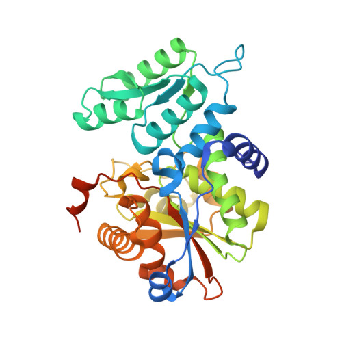
Tyrosine 121 moves revealing a ligandable pocket that couples catalysis to ATP-binding in serine racemase.

Koulouris, C.R.Gardiner, S.E.Harris, T.K.Elvers, K.T.Mark Roe, S.Gillespie, J.A.Ward, S.E.Grubisha, O.Nicholls, R.A.Atack, J.R.Bax, B.D.

(2022) Commun Biol 5: 346-346

  • DOI: https://doi.org/10.1038/s42003-022-03264-5
  • Primary Citation Related Structures: 
    6ZSP, 6ZUJ, 7NBC, 7NBD, 7NBF, 7NBG, 7NBH

  • PubMed Abstract: 

    Human serine racemase (hSR) catalyses racemisation of L-serine to D-serine, the latter of which is a co-agonist of the NMDA subtype of glutamate receptors that are important in synaptic plasticity, learning and memory. In a 'closed' hSR structure containing the allosteric activator ATP, the inhibitor malonate is enclosed between the large and small domains while ATP is distal to the active site, residing at the dimer interface with the Tyr121 hydroxyl group contacting the α-phosphate of ATP. In contrast, in 'open' hSR structures, Tyr121 sits in the core of the small domain with its hydroxyl contacting the key catalytic residue Ser84. The ability to regulate SR activity by flipping Tyr121 from the core of the small domain to the dimer interface appears to have evolved in animals with a CNS. Multiple X-ray crystallographic enzyme-fragment structures show Tyr121 flipped out of its pocket in the core of the small domain. Data suggest that this ligandable pocket could be targeted by molecules that inhibit enzyme activity.


  • Organizational Affiliation
    • Sussex Drug Discovery Centre, University of Sussex, Brighton, BN1 9QG, UK.

Macromolecule Content 

  • Total Structure Weight: 153.47 kDa 
  • Atom Count: 11,331 
  • Modeled Residue Count: 1,275 
  • Deposited Residue Count: 1,384 
  • Unique protein chains: 1

Macromolecules

Find similar proteins by:|  3D Structure
Entity ID: 1
MoleculeChains  Sequence LengthOrganismDetailsImage
Serine racemaseA [auth AAA],
B [auth BBB],
C [auth CCC],
D [auth DDD]
346Homo sapiensMutation(s): 2 
Gene Names: SRR
EC: 5.1.1.18 (PDB Primary Data), 4.3.1.18 (PDB Primary Data), 4.3.1.17 (PDB Primary Data)
UniProt & NIH Common Fund Data Resources
Find proteins for Q9GZT4 (Homo sapiens)
Explore Q9GZT4 
Go to UniProtKB:  Q9GZT4
PHAROS:  Q9GZT4
GTEx:  ENSG00000167720 
Entity Groups
Sequence Clusters30% Identity50% Identity70% Identity90% Identity95% Identity100% Identity
UniProt GroupQ9GZT4
Sequence Annotations
Expand
Reference Sequence

Small Molecules

Ligands 10 Unique
IDChains Name / Formula / InChI Key2D Diagram3D Interactions
PLP

Query on PLP


Download:Ideal Coordinates CCD File
E [auth AAA],
GA [auth DDD],
P [auth BBB],
X [auth CCC]
PYRIDOXAL-5'-PHOSPHATE
C8 H10 N O6 P
NGVDGCNFYWLIFO-UHFFFAOYSA-N
T6J
(Subject of Investigation/LOI)

Query on T6J


Download:Ideal Coordinates CCD File
AA [auth CCC],
KA [auth DDD],
M [auth AAA],
U [auth BBB]
2-[(methylsulfonyl)methyl]-1H-benzimidazole
C9 H10 N2 O2 S
WCWTYTQSZZWNEW-UHFFFAOYSA-N
PGE

Query on PGE


Download:Ideal Coordinates CCD File
DA [auth CCC]TRIETHYLENE GLYCOL
C6 H14 O4
ZIBGPFATKBEMQZ-UHFFFAOYSA-N
GOL

Query on GOL


Download:Ideal Coordinates CCD File
BA [auth CCC]
EA [auth CCC]
F [auth AAA]
G [auth AAA]
H [auth AAA]
BA [auth CCC],
EA [auth CCC],
F [auth AAA],
G [auth AAA],
H [auth AAA],
L [auth AAA],
Q [auth BBB],
R [auth BBB],
Y [auth CCC]
GLYCEROL
C3 H8 O3
PEDCQBHIVMGVHV-UHFFFAOYSA-N
DMS

Query on DMS


Download:Ideal Coordinates CCD File
K [auth AAA]DIMETHYL SULFOXIDE
C2 H6 O S
IAZDPXIOMUYVGZ-UHFFFAOYSA-N
EDO

Query on EDO


Download:Ideal Coordinates CCD File
CA [auth CCC],
LA [auth DDD],
MA [auth DDD],
NA [auth DDD],
V [auth BBB]
1,2-ETHANEDIOL
C2 H6 O2
LYCAIKOWRPUZTN-UHFFFAOYSA-N
CA

Query on CA


Download:Ideal Coordinates CCD File
HA [auth DDD]
I [auth AAA]
N [auth AAA]
S [auth BBB]
T [auth BBB]
HA [auth DDD],
I [auth AAA],
N [auth AAA],
S [auth BBB],
T [auth BBB],
Z [auth CCC]
CALCIUM ION
Ca
BHPQYMZQTOCNFJ-UHFFFAOYSA-N
CL

Query on CL


Download:Ideal Coordinates CCD File
FA [auth CCC],
PA [auth DDD]
CHLORIDE ION
Cl
VEXZGXHMUGYJMC-UHFFFAOYSA-M
MG

Query on MG


Download:Ideal Coordinates CCD File
IA [auth DDD],
J [auth AAA]
MAGNESIUM ION
Mg
JLVVSXFLKOJNIY-UHFFFAOYSA-N
NA

Query on NA


Download:Ideal Coordinates CCD File
JA [auth DDD],
O [auth AAA],
OA [auth DDD],
W [auth BBB]
SODIUM ION
Na
FKNQFGJONOIPTF-UHFFFAOYSA-N

Experimental Data & Validation

Experimental Data

  • Method: X-RAY DIFFRACTION
  • Resolution: 1.60 Å
  • R-Value Free:  0.198 (Depositor), 0.190 (DCC) 
  • R-Value Work:  0.172 (Depositor) 
Space Group: P 1 21 1
Unit Cell:
Length ( Å )Angle ( ˚ )
a = 48.112α = 90
b = 154.937β = 97.957
c = 85.539γ = 90
Software Package:
Software NamePurpose
REFMACrefinement
DIALSdata reduction
Aimlessdata scaling
MOLREPphasing

Structure Validation

View Full Validation Report


Ligand Structure Quality Assessment 


Entry History 

& Funding Information

Deposition Data


Funding OrganizationLocationGrant Number
Medical Research Council (MRC, United Kingdom)United Kingdom--

Revision History  (Full details and data files)

  • Version 1.0: 2021-03-03
    Type: Initial release
  • Version 1.1: 2022-04-27
    Changes: Database references
  • Version 1.2: 2022-05-04
    Changes: Structure summary
  • Version 1.3: 2024-01-31
    Changes: Data collection, Database references, Derived calculations, Refinement description