7NAY | pdb_00007nay

Crystal structure of lactate dehydrogenase from Selenomonas ruminantium with NADH.


Experimental Data Snapshot

  • Method: X-RAY DIFFRACTION
  • Resolution: 1.84 Å
  • R-Value Free: 
    0.188 (Depositor), 0.196 (DCC) 
  • R-Value Work: 
    0.145 (Depositor), 0.158 (DCC) 
  • R-Value Observed: 
    0.149 (Depositor) 

Starting Model: experimental
View more details

wwPDB Validation 3D Report Full Report

Validation slider image for 7NAY

Ligand Structure Quality Assessment 


This is version 1.2 of the entry. See complete history

Literature

Biochemical, structural and dynamical characterizations of the lactate dehydrogenase from Selenomonas ruminantium provide information about an intermediate evolutionary step prior to complete allosteric regulation acquisition in the super family of lactate and malate dehydrogenases.

Bertrand, Q.Coquille, S.Iorio, A.Sterpone, F.Madern, D.

(2023) J Struct Biol 215: 108039-108039

  • DOI: https://doi.org/10.1016/j.jsb.2023.108039
  • Primary Citation Related Structures: 
    7NAY, 8Q3C

  • PubMed Abstract: 

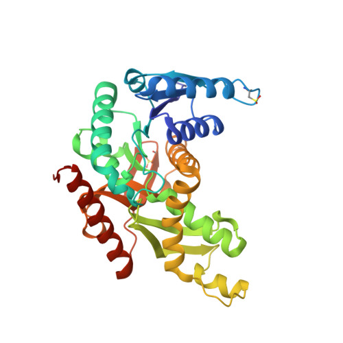
    In this work, we investigated the lactate dehydrogenase (LDH) from Selenomonas ruminantium (S. rum), an enzyme that differs at key amino acid positions from canonical allosteric LDHs. The wild type (Wt) of this enzyme recognises pyuvate as all LDHs. However, introducing a single point mutation in the active site loop (I85R) allows S. Rum LDH to recognize the oxaloacetate substrate as a typical malate dehydrogenase (MalDH), whilst maintaining homotropic activation as an LDH. We report the tertiary structure of the Wt and I85RLDH mutant. The Wt S. rum enzyme structure binds NADH and malonate, whilst also resembling the typical compact R-active state of canonical LDHs. The structure of the mutant with I85R was solved in the Apo State (without ligand), and shows no large conformational reorganization such as that observed with canonical allosteric LDHs in Apo state. This is due to a local structural feature typical of S. rum LDH that prevents large-scale conformational reorganization. The S. rum LDH was also studied using Molecular Dynamics simulations, probing specific local deformations of the active site that allow the S. rum LDH to sample the T-inactive state. We propose that, with respect to the LDH/MalDH superfamily, the S. rum enzyme possesses a specificstructural and dynamical way to ensure homotropic activation.


  • Organizational Affiliation
    • Univ. Grenoble Alpes, CEA, CNRS, IBS, 38000 Grenoble, France; Laboratory of Biomolecular Research, Biology and Chemistry Division, Paul Scherrer Institut, Villigen, Switzerland.

Macromolecule Content 

  • Total Structure Weight: 37.71 kDa 
  • Atom Count: 2,978 
  • Modeled Residue Count: 322 
  • Deposited Residue Count: 324 
  • Unique protein chains: 1

Macromolecules

Find similar proteins by:|  3D Structure
Entity ID: 1
MoleculeChains  Sequence LengthOrganismDetailsImage
L-lactate dehydrogenase324Selenomonas ruminantiumMutation(s): 0 
Gene Names: ldhldhL
EC: 1.1.1.27
UniProt
Find proteins for Q9EVR0 (Selenomonas ruminantium)
Explore Q9EVR0 
Go to UniProtKB:  Q9EVR0
Entity Groups
Sequence Clusters30% Identity50% Identity70% Identity90% Identity95% Identity100% Identity
UniProt GroupQ9EVR0
Sequence Annotations
Expand
Reference Sequence

Small Molecules

Ligands 5 Unique
IDChains Name / Formula / InChI Key2D Diagram3D Interactions
NAD

Query on NAD


Download:Ideal Coordinates CCD File
B [auth A]NICOTINAMIDE-ADENINE-DINUCLEOTIDE
C21 H27 N7 O14 P2
BAWFJGJZGIEFAR-NNYOXOHSSA-N
1PE

Query on 1PE


Download:Ideal Coordinates CCD File
J [auth A],
L [auth A]
PENTAETHYLENE GLYCOL
C10 H22 O6
JLFNLZLINWHATN-UHFFFAOYSA-N
MLA

Query on MLA


Download:Ideal Coordinates CCD File
C [auth A],
D [auth A]
MALONIC ACID
C3 H4 O4
OFOBLEOULBTSOW-UHFFFAOYSA-N
EDO

Query on EDO


Download:Ideal Coordinates CCD File
F [auth A],
G [auth A],
H [auth A],
I [auth A],
M [auth A]
1,2-ETHANEDIOL
C2 H6 O2
LYCAIKOWRPUZTN-UHFFFAOYSA-N
BO3

Query on BO3


Download:Ideal Coordinates CCD File
E [auth A],
K [auth A]
BORIC ACID
B H3 O3
KGBXLFKZBHKPEV-UHFFFAOYSA-N
Modified Residues  2 Unique
IDChains TypeFormula2D DiagramParent
CSD
Query on CSD
A
L-PEPTIDE LINKINGC3 H7 N O4 SCYS
OCS
Query on OCS
A
L-PEPTIDE LINKINGC3 H7 N O5 SCYS

Experimental Data & Validation

Experimental Data

  • Method: X-RAY DIFFRACTION
  • Resolution: 1.84 Å
  • R-Value Free:  0.188 (Depositor), 0.196 (DCC) 
  • R-Value Work:  0.145 (Depositor), 0.158 (DCC) 
  • R-Value Observed: 0.149 (Depositor) 
Space Group: I 2 2 2
Unit Cell:
Length ( Å )Angle ( ˚ )
a = 62.75α = 90
b = 104.26β = 90
c = 110.94γ = 90
Software Package:
Software NamePurpose
REFMACrefinement
PDB_EXTRACTdata extraction
XDSdata reduction
XDSdata scaling
PHASERphasing

Structure Validation

View Full Validation Report


Ligand Structure Quality Assessment 


Entry History 

& Funding Information

Deposition Data


Funding OrganizationLocationGrant Number
Agence Nationale de la Recherche (ANR)France--

Revision History  (Full details and data files)

  • Version 1.0: 2021-02-10
    Type: Initial release
  • Version 1.1: 2024-01-31
    Changes: Data collection, Database references, Refinement description
  • Version 1.2: 2024-03-20
    Changes: Database references