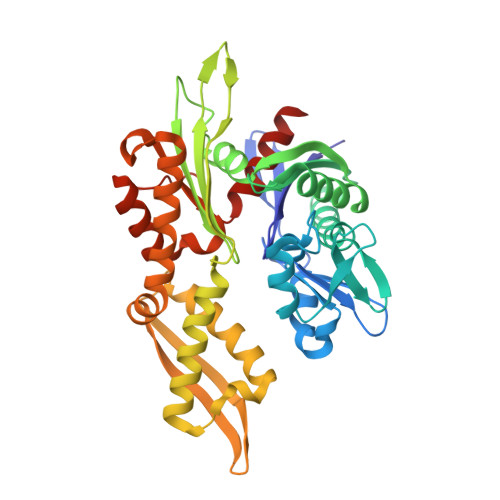
7N46

ADP-binding state of the nucleotide-binding domain of Hsp70 DnaK


Experimental Data Snapshot

  • Method: X-RAY DIFFRACTION
  • Resolution: 2.21 Å
  • R-Value Free: 0.248 
  • R-Value Work: 0.216 
  • R-Value Observed: 0.217 

Starting Model: experimental
View more details

wwPDB Validation   3D Report Full Report


Ligand Structure Quality Assessment 


This is version 1.2 of the entry. See complete history


Literature

Conformational equilibria in allosteric control of Hsp70 chaperones.

Wang, W.Liu, Q.Liu, Q.Hendrickson, W.A.

(2021) Mol Cell 81: 3919-3933.e7

  • DOI: https://doi.org/10.1016/j.molcel.2021.07.039
  • Primary Citation of Related Structures:  
    7KO2, 7KRT, 7KRU, 7KRV, 7KRW, 7N46, 7RAX

  • PubMed Abstract: 

    Heat-shock proteins of 70 kDa (Hsp70s) are vital for all life and are notably important in protein folding. Hsp70s use ATP binding and hydrolysis at a nucleotide-binding domain (NBD) to control the binding and release of client polypeptides at a substrate-binding domain (SBD); however, the mechanistic basis for this allostery has been elusive. Here, we first characterize biochemical properties of selected domain-interface mutants in bacterial Hsp70 DnaK. We then develop a theoretical model for allosteric equilibria among Hsp70 conformational states to explain the observations: a restraining state, Hsp70 R -ATP, restricts ATP hydrolysis and binds peptides poorly, whereas a stimulating state, Hsp70 S -ATP, hydrolyzes ATP rapidly and has high intrinsic substrate affinity but rapid binding kinetics. We support this model for allosteric regulation with DnaK structures obtained in the postulated stimulating state S with biochemical tests of the S-state interface and with improved peptide-binding-site definition in an R-state structure.


  • Organizational Affiliation

    Department of Biochemistry and Molecular Biophysics, Columbia University, New York, NY 10032, USA; Department of Biological Sciences, Columbia University, New York, NY 10027, USA.


Macromolecules
Find similar proteins by:  (by identity cutoff)  |  3D Structure
Entity ID: 1
MoleculeChains Sequence LengthOrganismDetailsImage
Chaperone protein DnaK386Escherichia coli K-12Mutation(s): 0 
Gene Names: dnaKFAZ83_07380
Entity Groups  
Sequence Clusters30% Identity50% Identity70% Identity90% Identity95% Identity100% Identity
Sequence Annotations
Expand
  • Reference Sequence
Experimental Data & Validation

Experimental Data

  • Method: X-RAY DIFFRACTION
  • Resolution: 2.21 Å
  • R-Value Free: 0.248 
  • R-Value Work: 0.216 
  • R-Value Observed: 0.217 
  • Space Group: P 65 2 2
Unit Cell:
Length ( Å )Angle ( ˚ )
a = 101.646α = 90
b = 101.646β = 90
c = 153.444γ = 120
Software Package:
Software NamePurpose
PHENIXrefinement
Aimlessdata scaling
PDB_EXTRACTdata extraction
XDSdata reduction
PHASERphasing

Structure Validation

View Full Validation Report


Ligand Structure Quality Assessment 


Entry History & Funding Information

Deposition Data


Funding OrganizationLocationGrant Number
National Institutes of Health/National Institute of General Medical Sciences (NIH/NIGMS)United StatesGM107462

Revision History  (Full details and data files)

  • Version 1.0: 2023-07-05
    Type: Initial release
  • Version 1.1: 2023-10-25
    Changes: Data collection, Refinement description
  • Version 1.2: 2024-07-17
    Changes: Database references