7MWF

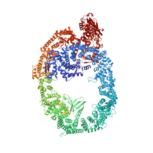
HUWE1 in map with focus on interface

  • Classification: TRANSFERASE
  • Organism(s): Homo sapiens
  • Expression System: Homo sapiens
  • Mutation(s): No 

  • Deposited: 2021-05-16 Released: 2021-07-28 
  • Deposition Author(s): Hunkeler, M., Fischer, E.S.
  • Funding Organization(s): National Institutes of Health/National Cancer Institute (NIH/NCI), Swiss National Science Foundation, Other private

Experimental Data Snapshot

  • Method: ELECTRON MICROSCOPY
  • Resolution: 3.30 Å
  • Aggregation State: PARTICLE 
  • Reconstruction Method: SINGLE PARTICLE 

wwPDB Validation   3D Report Full Report


This is version 1.3 of the entry. See complete history


Literature

Solenoid architecture of HUWE1 contributes to ligase activity and substrate recognition.

Hunkeler, M.Jin, C.Y.Ma, M.W.Monda, J.K.Overwijn, D.Bennett, E.J.Fischer, E.S.

(2021) Mol Cell 81: 3468

  • DOI: https://doi.org/10.1016/j.molcel.2021.06.032
  • Primary Citation of Related Structures:  
    7JQ9, 7MOP, 7MWD, 7MWE, 7MWF

  • PubMed Abstract: 

    HECT ubiquitin ligases play essential roles in metazoan development and physiology. The HECT ligase HUWE1 is central to the cellular stress response by mediating degradation of key death or survival factors, including Mcl1, p53, DDIT4, and Myc. Although mutations in HUWE1 and related HECT ligases are widely implicated in human disease, our molecular understanding remains limited. Here we present a comprehensive investigation of full-length HUWE1, deepening our understanding of this class of enzymes. The N-terminal ∼3,900 amino acids of HUWE1 are indispensable for proper ligase function, and our cryo-EM structures of HUWE1 offer a complete molecular picture of this large HECT ubiquitin ligase. HUWE1 forms an alpha solenoid-shaped assembly with a central pore decorated with protein interaction modules. Structures of HUWE1 variants linked to neurodevelopmental disorders as well as of HUWE1 bound to a model substrate link the functions of this essential enzyme to its three-dimensional organization.


  • Organizational Affiliation

    Department of Cancer Biology, Dana-Farber Cancer Institute, Boston, MA 02215, USA; Department of Biological Chemistry and Molecular Pharmacology, Harvard Medical School, Boston, MA 02115, USA.


Macromolecules
Find similar proteins by:  (by identity cutoff)  |  3D Structure
Entity ID: 1
MoleculeChains Sequence LengthOrganismDetailsImage
E3 ubiquitin-protein ligase HUWE14,411Homo sapiensMutation(s): 0 
Gene Names: HUWE1KIAA0312KIAA1578UREB1HSPC272
EC: 2.3.2.26
UniProt & NIH Common Fund Data Resources
Find proteins for Q7Z6Z7 (Homo sapiens)
Explore Q7Z6Z7 
Go to UniProtKB:  Q7Z6Z7
PHAROS:  Q7Z6Z7
GTEx:  ENSG00000086758 
Entity Groups  
Sequence Clusters30% Identity50% Identity70% Identity90% Identity95% Identity100% Identity
UniProt GroupQ7Z6Z7
Sequence Annotations
Expand
  • Reference Sequence
Experimental Data & Validation

Experimental Data

  • Method: ELECTRON MICROSCOPY
  • Resolution: 3.30 Å
  • Aggregation State: PARTICLE 
  • Reconstruction Method: SINGLE PARTICLE 
EM Software:
TaskSoftware PackageVersion
RECONSTRUCTIONRELION
MODEL REFINEMENTPHENIX
MODEL REFINEMENTISOLDE

Structure Validation

View Full Validation Report


Entry History & Funding Information

Deposition Data


Funding OrganizationLocationGrant Number
National Institutes of Health/National Cancer Institute (NIH/NCI)United StatesR01CA2144608
National Institutes of Health/National Cancer Institute (NIH/NCI)United StatesR01CA218278
Swiss National Science FoundationSwitzerland174331
Swiss National Science FoundationSwitzerland191053
Other privateUnited StatesDRR-50-18

Revision History  (Full details and data files)

  • Version 1.0: 2021-07-28
    Type: Initial release
  • Version 1.1: 2021-08-25
    Changes: Database references
  • Version 1.2: 2021-09-15
    Changes: Database references
  • Version 1.3: 2024-05-29
    Changes: Data collection