7MFR

Crystal Structure of a Fab fragment bound to peptide GGM


Experimental Data Snapshot

  • Method: X-RAY DIFFRACTION
  • Resolution: 2.85 Å
  • R-Value Free: 0.260 
  • R-Value Work: 0.211 
  • R-Value Observed: 0.213 

wwPDB Validation   3D Report Full Report


This is version 1.3 of the entry. See complete history


Literature

Antibody toolkit reveals N-terminally ubiquitinated substrates of UBE2W.

Davies, C.W.Vidal, S.E.Phu, L.Sudhamsu, J.Hinkle, T.B.Chan Rosenberg, S.Schumacher, F.R.Zeng, Y.J.Schwerdtfeger, C.Peterson, A.S.Lill, J.R.Rose, C.M.Shaw, A.S.Wertz, I.E.Kirkpatrick, D.S.Koerber, J.T.

(2021) Nat Commun 12: 4608-4608

  • DOI: https://doi.org/10.1038/s41467-021-24669-6
  • Primary Citation of Related Structures:  
    7MFR

  • PubMed Abstract: 

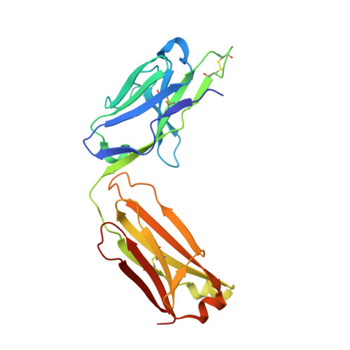
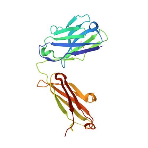
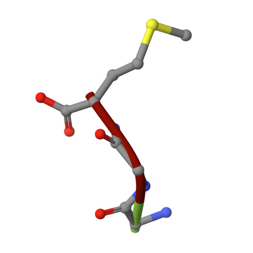
    The ubiquitin conjugating enzyme UBE2W catalyzes non-canonical ubiquitination on the N-termini of proteins, although its substrate repertoire remains unclear. To identify endogenous N-terminally-ubiquitinated substrates, we discover four monoclonal antibodies that selectively recognize tryptic peptides with an N-terminal diglycine remnant, corresponding to sites of N-terminal ubiquitination. Importantly, these antibodies do not recognize isopeptide-linked diglycine (ubiquitin) modifications on lysine. We solve the structure of one such antibody bound to a Gly-Gly-Met peptide to reveal the molecular basis for its selective recognition. We use these antibodies in conjunction with mass spectrometry proteomics to map N-terminal ubiquitination sites on endogenous substrates of UBE2W. These substrates include UCHL1 and UCHL5, where N-terminal ubiquitination distinctly alters deubiquitinase (DUB) activity. This work describes an antibody toolkit for enrichment and global profiling of endogenous N-terminal ubiquitination sites, while revealing functionally relevant substrates of UBE2W.


  • Organizational Affiliation

    Department of Antibody Engineering, Genentech, Inc., South San Francisco, CA, USA.


Macromolecules
Find similar proteins by:  (by identity cutoff)  |  3D Structure
Entity ID: 1
MoleculeChains Sequence LengthOrganismDetailsImage
Antibody fragment - Light Chain of fabA,
C [auth X]
220Oryctolagus cuniculusMutation(s): 0 
Entity Groups  
Sequence Clusters30% Identity50% Identity70% Identity90% Identity95% Identity100% Identity
Sequence Annotations
Expand
  • Reference Sequence
Find similar proteins by:  (by identity cutoff)  |  3D Structure
Entity ID: 2
MoleculeChains Sequence LengthOrganismDetailsImage
Antibody fragment - Heavy Chain of fabB,
D [auth Y]
217Oryctolagus cuniculusMutation(s): 0 
Entity Groups  
Sequence Clusters30% Identity50% Identity70% Identity90% Identity95% Identity100% Identity
Sequence Annotations
Expand
  • Reference Sequence

Find similar proteins by:  Sequence   |   3D Structure  

Entity ID: 3
MoleculeChains Sequence LengthOrganismDetailsImage
GLY-GLY-METE [auth P],
F [auth Q]
3Homo sapiensMutation(s): 0 
Sequence Annotations
Expand
  • Reference Sequence
Experimental Data & Validation

Experimental Data

  • Method: X-RAY DIFFRACTION
  • Resolution: 2.85 Å
  • R-Value Free: 0.260 
  • R-Value Work: 0.211 
  • R-Value Observed: 0.213 
  • Space Group: P 43 21 2
Unit Cell:
Length ( Å )Angle ( ˚ )
a = 163.797α = 90
b = 163.797β = 90
c = 127.471γ = 90
Software Package:
Software NamePurpose
SCALEPACKdata scaling
PHENIXrefinement
PDB_EXTRACTdata extraction
HKL-2000data reduction
MOLREPphasing

Structure Validation

View Full Validation Report


Entry History 

Deposition Data

  • Released Date: 2021-06-16 
  • Deposition Author(s): Sudhamsu, J.

Revision History  (Full details and data files)

  • Version 1.0: 2021-06-16
    Type: Initial release
  • Version 1.1: 2021-12-29
    Changes: Database references
  • Version 1.2: 2022-01-05
    Changes: Database references
  • Version 1.3: 2023-10-18
    Changes: Data collection, Refinement description