7MFH | pdb_00007mfh

Crystal structure of BIO-32546 bound mouse Autotaxin


Experimental Data Snapshot

  • Method: X-RAY DIFFRACTION
  • Resolution: 2.30 Å
  • R-Value Free: 
    0.268 (Depositor), 0.270 (DCC) 
  • R-Value Work: 
    0.215 (Depositor), 0.215 (DCC) 
  • R-Value Observed: 
    0.218 (Depositor) 

Starting Model: experimental
View more details

wwPDB Validation 3D Report Full Report

Validation slider image for 7MFH

Ligand Structure Quality Assessment 


This is version 1.2 of the entry. See complete history

Literature

Discovery of Potent Selective Nonzinc Binding Autotaxin Inhibitor BIO-32546.

Ma, B.Zhang, L.Sun, L.Xin, Z.Kumaravel, G.Marcotte, D.Chodaparambil, J.V.Wang, Q.Wehr, A.Jing, J.Hong, V.S.Wang, T.Huang, C.Shao, Z.Mi, S.

(2021) ACS Med Chem Lett 12: 1124-1129

  • DOI: https://doi.org/10.1021/acsmedchemlett.1c00211
  • Primary Citation Related Structures: 
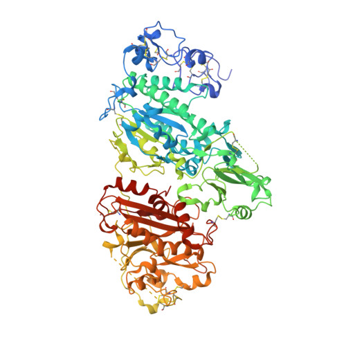
    7MFH

  • PubMed Abstract: 

    Autotaxin (ATX) is a lysophospholipase D that is the main enzyme responsible for generating LPA in body fluids. Although ATX was isolated from a conditioned medium of melanoma cells, later it was discovered to play a critical role in vascular and neuronal development. ATX has also been implicated in primary brain tumor, fibrosis, and rheumatoid arthritis, as well as neurological diseases such as multiple sclerosis, Alzheimer's disease, and neuropathic pain. As ATX and LPA levels are increased upon neuronal injury, a selective ATX inhibitor could provide a new approach to treat neuropathic pain. Herein we describe the discovery of a novel series of nonzinc binding reversible ATX inhibitors, particularly a potent, selective, orally bioavailable, brain-penetrable tool compound BIO-32546, as well as its synthesis, X-ray cocrystal structure, pharmacokinetics, and in vivo efficacy.


  • Organizational Affiliation
    • Medicinal Chemistry, Physical Biochemistry, Drug Metabolism & Pharmacokinetics, Discovery Bioassay, Neurology, Biogen Inc., 225 Binney St, Cambridge, Massachusetts 02142, United States.

Macromolecule Content 

  • Total Structure Weight: 99.45 kDa 
  • Atom Count: 6,618 
  • Modeled Residue Count: 776 
  • Deposited Residue Count: 832 
  • Unique protein chains: 1

Macromolecules

Find similar proteins by:|  3D Structure
Entity ID: 1
MoleculeChains  Sequence LengthOrganismDetailsImage
Ectonucleotide pyrophosphatase/phosphodiesterase family member 2832Mus musculusMutation(s): 0 
Gene Names: Enpp2Npps2Pdnp2
EC: 3.1.4.39 (PDB Primary Data), 3.1.4.4 (UniProt)
UniProt
Find proteins for Q9R1E6 (Mus musculus)
Explore Q9R1E6 
Go to UniProtKB:  Q9R1E6
Entity Groups
Sequence Clusters30% Identity50% Identity70% Identity90% Identity95% Identity100% Identity
UniProt GroupQ9R1E6
Glycosylation
Glycosylation Sites: 2Go to GlyGen: Q9R1E6-1
Sequence Annotations
Expand
Reference Sequence

Oligosaccharides

Help  
Entity ID: 2
MoleculeChains Length2D Diagram GlycosylationD Interactions
alpha-D-mannopyranose-(1-2)-alpha-D-mannopyranose-(1-3)-alpha-D-mannopyranose-(1-6)-beta-D-mannopyranose-(1-4)-2-acetamido-2-deoxy-beta-D-glucopyranose-(1-4)-2-acetamido-2-deoxy-beta-D-glucopyranose
B
6N-Glycosylation
Glycosylation Resources
GlyTouCan: G47410OF
GlyCosmos: G47410OF
GlyGen: G47410OF
Entity ID: 3
MoleculeChains Length2D Diagram GlycosylationD Interactions
2-acetamido-2-deoxy-beta-D-glucopyranose-(1-4)-2-acetamido-2-deoxy-beta-D-glucopyranose
C
2N-Glycosylation
Glycosylation Resources
GlyTouCan: G42666HT
GlyCosmos: G42666HT
GlyGen: G42666HT

Small Molecules

Ligands 5 Unique
IDChains Name / Formula / InChI Key2D Diagram3D Interactions
ZF7
(Subject of Investigation/LOI)

Query on ZF7


Download:Ideal Coordinates CCD File
D [auth A](1R,3S,5S)-8-{(1S)-1-[8-(trifluoromethyl)-7-{[(1s,4R)-4-(trifluoromethyl)cyclohexyl]oxy}naphthalen-2-yl]ethyl}-8-azabicyclo[3.2.1]octane-3-carboxylic acid
C28 H31 F6 N O3
PZASAAIJIFDWSB-CUWQMCGTSA-N
IOD

Query on IOD


Download:Ideal Coordinates CCD File
E [auth A]IODIDE ION
I
XMBWDFGMSWQBCA-UHFFFAOYSA-M
PEG

Query on PEG


Download:Ideal Coordinates CCD File
M [auth A]DI(HYDROXYETHYL)ETHER
C4 H10 O3
MTHSVFCYNBDYFN-UHFFFAOYSA-N
ZN

Query on ZN


Download:Ideal Coordinates CCD File
G [auth A]
O [auth A]
P [auth A]
V [auth A]
W [auth A]
G [auth A],
O [auth A],
P [auth A],
V [auth A],
W [auth A],
X [auth A],
Y [auth A]
ZINC ION
Zn
PTFCDOFLOPIGGS-UHFFFAOYSA-N
EDO

Query on EDO


Download:Ideal Coordinates CCD File
AA [auth A]
F [auth A]
H [auth A]
I [auth A]
J [auth A]
AA [auth A],
F [auth A],
H [auth A],
I [auth A],
J [auth A],
K [auth A],
L [auth A],
N [auth A],
Q [auth A],
R [auth A],
S [auth A],
T [auth A],
U [auth A],
Z [auth A]
1,2-ETHANEDIOL
C2 H6 O2
LYCAIKOWRPUZTN-UHFFFAOYSA-N

Experimental Data & Validation

Experimental Data

  • Method: X-RAY DIFFRACTION
  • Resolution: 2.30 Å
  • R-Value Free:  0.268 (Depositor), 0.270 (DCC) 
  • R-Value Work:  0.215 (Depositor), 0.215 (DCC) 
  • R-Value Observed: 0.218 (Depositor) 
Space Group: P 1
Unit Cell:
Length ( Å )Angle ( ˚ )
a = 61.02α = 89.071
b = 61.62β = 74.202
c = 67.32γ = 79.877
Software Package:
Software NamePurpose
PHENIXrefinement
XDSdata reduction
Aimlessdata scaling
PHENIXphasing

Structure Validation

View Full Validation Report


Ligand Structure Quality Assessment 


Entry History 

Deposition Data

Revision History  (Full details and data files)

  • Version 1.0: 2022-04-13
    Type: Initial release
  • Version 1.1: 2023-10-18
    Changes: Data collection, Refinement description
  • Version 1.2: 2024-11-20
    Changes: Structure summary