7MF3

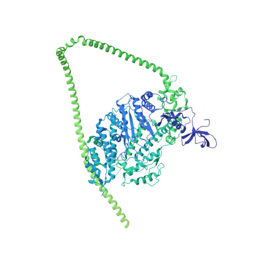
Structure of the autoinhibited state of smooth muscle myosin-2


Experimental Data Snapshot

  • Method: ELECTRON MICROSCOPY
  • Resolution: 3.40 Å
  • Aggregation State: TISSUE 
  • Reconstruction Method: SINGLE PARTICLE 

wwPDB Validation   3D Report Full Report


This is version 1.0 of the entry. See complete history


Literature

Cryo-EM structure of the autoinhibited state of myosin-2.

Heissler, S.M.Arora, A.S.Billington, N.Sellers, J.R.Chinthalapudi, K.

(2021) Sci Adv 7: eabk3273-eabk3273

  • DOI: https://doi.org/10.1126/sciadv.abk3273
  • Primary Citation of Related Structures:  
    7MF3

  • PubMed Abstract: 

    We solved the near-atomic resolution structure of smooth muscle myosin-2 in the autoinhibited state (10 S ) using single-particle cryo–electron microscopy. The 3.4-Å structure reveals the precise molecular architecture of 10 S and the structural basis for myosin-2 regulation. We reveal the position of the phosphorylation sites that control myosin autoinhibition and activation by phosphorylation of the regulatory light chain. Further, we present a previously unidentified conformational state in myosin-2 that traps ADP and P i produced by the hydrolysis of ATP in the active site. This noncanonical state represents a branch of the myosin enzyme cycle and explains the autoinhibition of the enzyme function of 10 S along with its reduced affinity for actin. Together, our structure defines the molecular mechanisms that drive 10 S formation, stabilization, and relief by phosphorylation of the regulatory light chain.


  • Organizational Affiliation

    Department of Physiology and Cell Biology, Dorothy M. Davis Heart and Lung Research Institute, The Ohio State University College of Medicine, Columbus, OH, USA.


Macromolecules
Find similar proteins by:  (by identity cutoff)  |  3D Structure
Entity ID: 1
MoleculeChains Sequence LengthOrganismDetailsImage
Myosin-11
A, B, G, H
1,978Gallus gallusMutation(s): 0 
UniProt
Find proteins for P10587 (Gallus gallus)
Explore P10587 
Go to UniProtKB:  P10587
Entity Groups  
Sequence Clusters30% Identity50% Identity70% Identity90% Identity95% Identity100% Identity
UniProt GroupP10587
Sequence Annotations
Expand
  • Reference Sequence
Find similar proteins by:  (by identity cutoff)  |  3D Structure
Entity ID: 2
MoleculeChains Sequence LengthOrganismDetailsImage
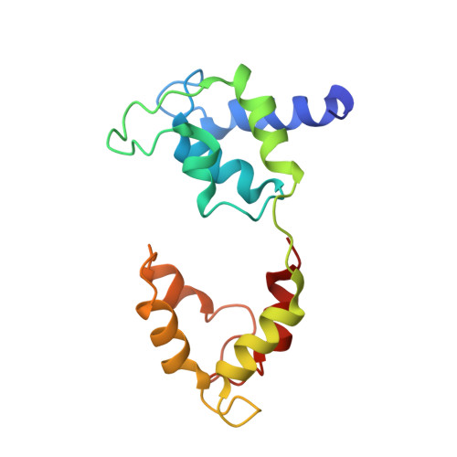
Myosin light polypeptide 6
C, F
150Gallus gallusMutation(s): 0 
UniProt
Find proteins for P02607 (Gallus gallus)
Explore P02607 
Go to UniProtKB:  P02607
Entity Groups  
Sequence Clusters30% Identity50% Identity70% Identity90% Identity95% Identity100% Identity
UniProt GroupP02607
Sequence Annotations
Expand
  • Reference Sequence
Find similar proteins by:  (by identity cutoff)  |  3D Structure
Entity ID: 3
MoleculeChains Sequence LengthOrganismDetailsImage
Myosin regulatory light chain 2, smooth muscle major isoform
D, E
171Gallus gallusMutation(s): 0 
UniProt
Find proteins for P02612 (Gallus gallus)
Explore P02612 
Go to UniProtKB:  P02612
Entity Groups  
Sequence Clusters30% Identity50% Identity70% Identity90% Identity95% Identity100% Identity
UniProt GroupP02612
Sequence Annotations
Expand
  • Reference Sequence
Small Molecules
Ligands 3 Unique
IDChains Name / Formula / InChI Key2D Diagram3D Interactions
ADP
Query on ADP

Download Ideal Coordinates CCD File 
I [auth A],
L [auth B]
ADENOSINE-5'-DIPHOSPHATE
C10 H15 N5 O10 P2
XTWYTFMLZFPYCI-KQYNXXCUSA-N
PO4
Query on PO4

Download Ideal Coordinates CCD File 
J [auth A],
M [auth B]
PHOSPHATE ION
O4 P
NBIIXXVUZAFLBC-UHFFFAOYSA-K
MG
Query on MG

Download Ideal Coordinates CCD File 
K [auth A],
N [auth B],
O [auth D],
P [auth E]
MAGNESIUM ION
Mg
JLVVSXFLKOJNIY-UHFFFAOYSA-N
Experimental Data & Validation

Experimental Data

  • Method: ELECTRON MICROSCOPY
  • Resolution: 3.40 Å
  • Aggregation State: TISSUE 
  • Reconstruction Method: SINGLE PARTICLE 
EM Software:
TaskSoftware PackageVersion
RECONSTRUCTIONcryoSPARC
MODEL REFINEMENTPHENIX

Structure Validation

View Full Validation Report


Entry History 

Deposition Data

Revision History  (Full details and data files)

  • Version 1.0: 2022-01-05
    Type: Initial release