Structural Insights into the Mechanism of Human T-cell Leukemia Virus Type 1 Gag Targeting to the Plasma Membrane for Assembly.
Herrmann, D., Zhou, L.W., Hanson, H.M., Willkomm, N.A., Mansky, L.M., Saad, J.S.(2021) J Mol Biology 433: 167161-167161
- PubMed: 34298060 Search on PubMedSearch on PubMed Central
- DOI: https://doi.org/10.1016/j.jmb.2021.167161
- Primary Citation Related Structures:
7M1W - PubMed Abstract:
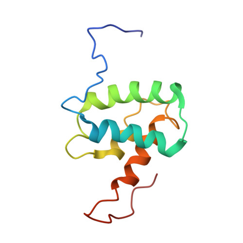
Retroviral Gag targeting to the plasma membrane (PM) for assembly is mediated by the N-terminal matrix (MA) domain. For many retroviruses, Gag-PM interaction is dependent on phosphatidylinositol 4,5-bisphosphate (PI(4,5)P 2 ). However, it has been shown that for human T-cell leukemia virus type 1 (HTLV-1), Gag binding to membranes is less dependent on PI(4,5)P 2 than HIV-1, suggesting that other factors may modulate Gag assembly. To elucidate the mechanism by which HTLV-1 Gag binds to the PM, we employed NMR techniques to determine the structure of unmyristoylated MA (myr(-)MA) and to characterize its interactions with lipids and liposomes. The MA structure consists of four α-helices and unstructured N- and C-termini. We show that myr(-)MA binds to PI(4,5)P 2 via the polar head and that binding to inositol phosphates (IPs) is significantly enhanced by increasing the number of phosphate groups on the inositol ring, indicating that the MA-IP binding is governed by charge-charge interactions. The IP binding site was mapped to a well-defined basic patch formed by lysine and arginine residues. Using an NMR-based liposome binding assay, we show that PI(4,5)P 2 and phosphatidylserine enhance myr(-)MA binding in a synergistic fashion. Confocal microscopy data revealed formation of puncta on the PM of Gag expressing cells. However, G2A-Gag mutant, lacking myristoylation, is diffuse and cytoplasmic. These results suggest that although myr(-)MA binds to membranes, myristoylation appears to be key for formation of HTLV-1 Gag puncta on the PM. Altogether, these findings advance our understanding of a key mechanism in retroviral assembly.
- Department of Microbiology, University of Alabama at Birmingham, Birmingham, AL 35294, USA.
Organizational Affiliation: