


Protein Model Building by the Use of a Constrained-Restrained Least-Squares Procedure
Herzberg, O., Sussman, J.L.(1983) J Appl Crystallogr 16: 144-150
Experimental Data Snapshot
wwPDB Validation 3D Report Full Report

(1983) J Appl Crystallogr 16: 144-150
Entity ID: 1 | |||||
|---|---|---|---|---|---|
| Molecule | Chains | Sequence Length | Organism | Details | Image |
| HEN EGG WHITE LYSOZYME | 129 | Gallus gallus | Mutation(s): 0 EC: 3.2.1.17 | 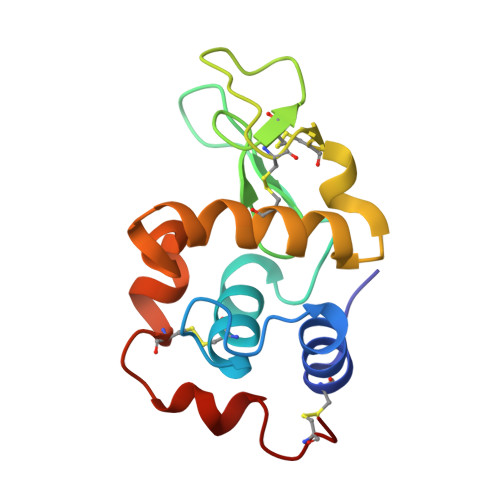 | |
UniProt | |||||
Entity Groups | |||||
| Sequence Clusters | 30% Identity50% Identity70% Identity90% Identity95% Identity100% Identity | ||||
| UniProt Group | P00698 | ||||
Sequence AnnotationsExpand | |||||
Reference Sequence | |||||
| Length ( Å ) | Angle ( ˚ ) |
|---|---|
| a = 27.4 | α = 88 |
| b = 31.9 | β = 108 |
| c = 34.4 | γ = 112 |
| Software Name | Purpose |
|---|---|
| CORELS | refinement |


